

Ultrasound guided synovial biopsy using portal and forceps
- PMID: 15550535
- PMCID: PMC1755510
- DOI: 10.1136/ard.2004.027409
Ultrasound guided synovial biopsy using portal and forceps
Abstract
Objective: To describe a new method for taking a synovial biopsy specimen under ultrasound guidance using portal and forceps.
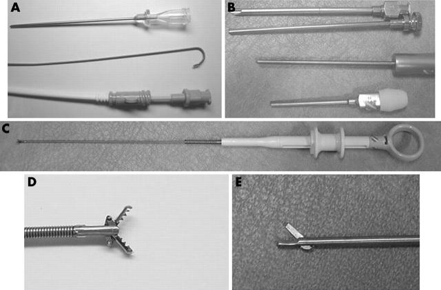
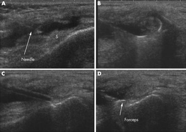
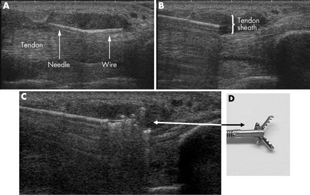
Methods: Percutaneous ultrasound guided biopsy was performed for 37 patients with mono- or polyarthritis as outpatients. A portal to a planned area was built using a needle, guiding wire, and dilators, through which forceps could be inserted and samples taken. Biopsy samples were taken from small and large joints, bursae, and tendon sheaths.
Results: Representative synovial tissue in adequate amounts for histopathological evaluation was obtained in 33/37 cases--a success rate of 89%. The biopsy procedures were well tolerated, but one complication of skin infection was encountered.
Conclusion: The new method of synovium biopsy under ultrasound guidance using sheath introducer set and flexible forceps can be performed on most joints and even bursae and tendon sheaths. The method gives sufficient samples for clinical work in most cases, but further work is needed before accepting this promising technique for scientific purposes.
Figures
References
Publication types
MeSH terms
LinkOut - more resources
Full Text Sources
Medical
