. 2004 Nov 30;101(48):16879-84.
doi: 10.1073/pnas.0406678101. Epub 2004 Nov 18.

Activation of the complement system generates antibacterial peptides

Affiliations

Activation of the complement system generates antibacterial peptides

Emma Andersson Nordahl et al. Proc Natl Acad Sci U S A. .

Abstract

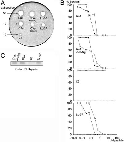
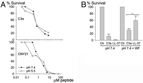
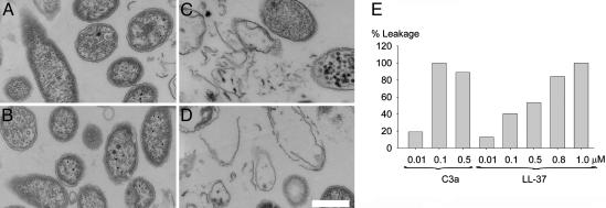
The complement system represents an evolutionary old and significant part of the innate immune system involved in protection against invading microorganisms. Here, we show that the anaphylatoxin C3a and its inactivated derivative C3a-desArg are antibacterial, demonstrating a previously unknown direct antimicrobial effect of complement activation. The C3a peptide, as well as functional epitopes in the sequence, efficiently killed the Gram-negative bacteria Escherichia coli, Pseudomonas aeruginosa, and the Gram-positive Enterococcus faecalis. In mice, a C3a-derived peptide suppressed infection by Gram-positive Streptococcus pyogenes bacteria. Fluorescence and electron microscopy demonstrated that C3a binds to and induces breaks in bacterial membranes. C3a was also found to induce membrane leakage of liposomes. These findings provide an interesting link between the complement system and antimicrobial peptides, which are two important branches of innate immunity.


Figures

Fig. 3.
Fig. 3.
Activities of synthetic C3a-derived peptides. (A) Structure of the anaphylatoxin C3a peptide, modified from Hugli 1989 (9). (B) The synthetic peptides used in this study are indicated in the sequence. (C) Viable-count assays were performed by using both E. faecalis 2374 (•), and P. aeruginosa 27.1 (○). We incubated 2 × 10⁶ cfu/ml bacteria in 50 μl with peptides at concentrations ranging 0.03–60 μM. For LGL5, the two graphs overlap. (D) Heparin-binding activity of the C3a-derived peptides. Peptides at indicated concentrations were applied to nitrocellulose membranes followed by incubation in PBS (containing 3% BSA) with iodinated (125I) heparin. LL-37 was used as positive control. Excess of unlabeled heparin (+) inhibited the binding of peptides to 125I-heparin. (E) Binding of TAMRA-labeled peptides to P. aeruginosa 27.1 and inhibition of binding by excess of heparin. Image 2 shows red fluorescence of bacteria (1 × 10⁷ ml-1) stained with TAMRA-conjugated peptides (10 μg/ml-1), and image 4 shows bacteria incubated with heparin and TAMRA-conjugated peptides. Images 2 and 4 were recorded by using identical instrument settings. Images 1 and 3 are the corresponding Nomarski images. (Scale bar, 10 μm.)
Fig. 5.
Fig. 5.
Antibacterial activities of CNY21 in vivo and generation of C3a-fragments ex vivo. (A) Space-filling model of CNY21 arranged as an α-helix. The arrows indicate positions of arginine residues that are replaced by serine residues in CNY21(S). (B) S. pyogenes bacteria were subjected to 10 μM CNY21(S) or CNY21 in 10 mM Tris (pH 7.4). At this dose, CNY21 was lethal to S. pyogenes whereas CNY21(S) had no effect; ***, P < 0.001 (n = 7). (C) C3a-derived peptides suppress bacterial dissemination to the spleen. Mice were i.p. injected with S. pyogenes bacteria, followed by i.p. injection with CNY21(S) (•) or CNY21 (○). The mice were killed 24 h after injection, and the total number of cfu in the spleen was determined for each mouse. Treatment with CNY21 yielded significantly lower bacterial numbers than treatment with CNY21(S). **, P = 0.006 (n = 14). Horizontal lines indicate the median value in each group. (D) Human WF (5 μl) was incubated at 37°C with lysed (0.3% Tween 20) neutrophils (corresponding to 9.4 × 10⁴ cells) for the indicated time periods. Western blot analysis identified cleavage products recognized by polyclonal antibodies against the C3a-derived peptide LGE27. No C3a fragments were detected in WF or neutrophils (polymorphonuclear leukocytes, PMN). Molecular-mass markers are indicated on the left. (E) Neutrophil elastase degrades C3a into several fragments. C3a (2 μg) was treated with human neutrophil elastase (40 milliunits) for indicated time periods at 37°C and analyzed by SDS/PAGE (16.5% Tris·tricine gel). For control, 0.5 μg of C3a was used. Molecular mass markers are indicated on the left. The fragment indicated with an arrow was analyzed by N-terminal sequencing.
Fig. 1.
Fig. 1.
Antibacterial and heparin-binding effects of C3a and C3a-desArg. (A) Peptides were tested in RDA in low-salt conditions. E. coli isolate 37.4 (4 × 10⁶ cfu) was inoculated in 0.1% TSB agarose gel. Each 4 mm-diameter well was loaded with 6 μl of peptide at the indicated concentration. The zones of clearance correspond to the inhibitory effect of each peptide after incubation at 37°C for 18–24 h. A negative control containing buffer (10 mM Tris, pH 7.4) was included in the well at the top left of the plate. This clear zone corresponds to the 4 mm well. (B) In viable-count assays antibacterial activities were seen against both E. faecalis isolate 2374 (•) and P. aeruginosa isolate 27.1 (○). We incubated 2 × 10⁶ cfu/ml bacteria in 50 μl with peptides at concentrations ranging 0.003–6 μM. For C3, the two graphs overlap. (C) C3a and C3a-desArg were both able to bind heparin, whereas only weak binding was seen with C3. C3a and C3a-desArg (2 μg), C3, and LL-37 (2 and 5 μg) were applied to nitrocellulose membranes. These membranes were then incubated in PBS (containing 3% BSA) with iodinated (125I) heparin. LL-37 was used as positive control.
Fig. 4.
Fig. 4.
Antibacterial activities of peptides under physiological conditions. (A) Viable-count analysis of C3a and CNY21 in different buffers; 10 mM Tris (pH 7.4) (•) and 10 mM Mes (pH 5.5) (○), both containing 0.15 M NaCl. P. aeruginosa 27.1 (2 × 10⁶ cfu/ml) were incubated in 50 μl with peptides at concentrations ranging 0.03–6 μM for C3a and 0.03–60 μM for CNY21. (B) P. aeruginosa bacteria were subjected to 10 μM C3a in 10 mM Tris (pH 7.4) containing 0.15 M NaCl in the presence or the absence of 20% WF. For comparison, LL-37 was used at the same concentration. ***, P < 0.001; **, P < 0.01.
Fig. 2.
Fig. 2.
C3a interacts with and generates breaks in bacterial plasma membranes. P. aeruginosa 27.1 was incubated with the holoprotein C3 and C3a peptides at 0.3 μM and analyzed with electron microscopy. Control (A), C3 (B), C3a (C), and C3a-desArg (D) are shown. (Scale bar, 0.5 μm.) (E) Effects of C3a and LL-37 on liposomes. The membrane permeabilizing effect was recorded by measuring fluorescence release of CF from liposomes. Values represents mean of double samples. A representative experiment of three is shown.

References

    1. Fearon, D. T. & Locksley, R. M. (1996) Science 272, 50-53. - PubMed
    1. Steiner, H., Hultmark, D., Engström, A., Bennich, H. & Boman, H. G. (1981) Nature 292, 246-248. - PubMed
    1. Schröder, J. M. & Harder, J. (1999) Int. J. Biochem. Cell Biol. 31, 645-651. - PubMed
    1. Boman, H. G. (2000) Immunol. Rev. 173, 5-16. - PubMed
    1. Zasloff, M. (2002) Nature 415, 389-395. - PubMed

Publication types