. 2004 Nov 18;32(20):6047-56.
doi: 10.1093/nar/gkh931. Print 2004.

The stability of Seeman JX DNA topoisomers of paranemic crossover (PX) molecules as a function of crossover number

Affiliations

The stability of Seeman JX DNA topoisomers of paranemic crossover (PX) molecules as a function of crossover number

Prabal K Maiti et al. Nucleic Acids Res. .

Abstract

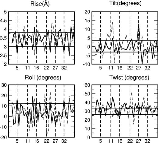
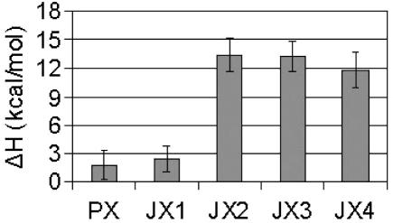
We use molecular dynamics simulations in explicit water and salt (Na+) to determine the effect of varying the number of crossover points on the structure and stability of the PX65 paranemic crossover DNA molecule and its JXM topoisomers (M denotes the number of missing crossover points), recently synthesized by the Seeman group at New York University. We find that PX65, with six crossover points, is the most stable, and that the stability decreases monotonically with the number of crossover points PX65 > JX1 > JX2 > JX3 > JX4, with 6, 5, 4, 3 and 2 crossover points, respectively. Thus, for PX65/JX1, the strain energy is approximately 3 kcal/mol/bp, while it is approximately 13 kcal/mol/bp for JX2, JX3 and JX4. Another measure of the stability is the change in the structure from the minimum energy structure to the equilibrium structure at 300 K, denoted as root-mean-square deviation in coordinates (CRMSD). We find that CRMSD is approximately 3.5 A for PX65, increases to 6 A for JX1 and increases to 10 A for JX2/JX3/JX4. As the number of crossover points decreases, the distance between the two double helical domains of the PX/JX molecules increases from approximately 20 A for PX65 to 23 A for JX4. This indicates that JX2, JX3 and JX4 are less likely to form, at least in with Na+. However, in all the cases, the two double helical domains have average helicoidal parameters similar to a typical B-DNA of similar length and base sequence.


Figures

Figure 1
Figure 1
The base pair sequences used in the generations of PX65, JX1, JX2, JX3 and JX4 crossover molecules.
Figure 2
Figure 2
Generation of PX and JX DNA by reciprocal exchange. This illustrates the consequences of performing a crossover at various positions. PX65 has six crossover points. JX1, JX2, JX3 and JX4 have 5, 4, 3 and 2 crossover points, respectively.
Figure 3
Figure 3
(a) Averaged dynamics structure for various PX molecules. Water molecules and counter ions are not shown for clarity. Note that with decreasing number of crossover points, the two double helical domains move further apart from each other. (b) Averaged dynamics structure for various PX molecules (side view). For clarity, water molecules and counter ions are not shown. With decreasing number of crossover points, there is significant bending of the two helical axis in the opposite direction leading to large writhing in the structure (see Table 4).
Figure 4
Figure 4
(a) Variation of the CRMSD of all atoms of various snapshots from the MD simulation run with respect to the starting minimized canonical structures. (b) RMSD with respect to average dynamics structure for different PX/JX molecules. The averaged structures were generated by averaging the coordinates for the last 1 ns of the 2 ns long MD runs. PX65/JX1 with CRMSD 3–5 Å is a stable molecule. Large CRMSD for JX2, JX3 and JX4 (8–10 Å) suggests that these structures will not retain their helical DNA structures and perhaps fall apart.
Figure 5
Figure 5
Distance between the center of mass of the two helices. With decreasing number of crossover points, the two helices move apart. The lowering of the distance for JX4 might be due to the large writhing during dynamics. The data has been averaged over the last 400 ps of the 2 ns long dynamics.
Figure 6
Figure 6
Average rise, tilt, roll and twist for PX motif. Solid line is for helix1 and broken line is for helix2. The vertical lines correspond to the crossover points. The horizontal solid lines give the upper bound and lower bound for the corresponding quantities expected for the helices in their B-DNA form(non-crossover form) during the dynamics. The data has been averaged over last 400 ps of the 3 ns long dynamics. In general, the two double helical domains in the crossover structure keep their B-DNA form quite well. However, at or near crossover points the helical parameters deviate significantly from the values expected in their B-DNA form.
Figure 7
Figure 7
Bending angle between every i-th and (i + 5)-th nucleotide for (a) helix1 and (b) helix2 for each of the PX/JX structures. There is no appreciable bending visible for PX65. However, with decreasing number of crossover points, increased bending occurs at or near each missing crossover point.
Figure 8
Figure 8
Strain energy for various PX/JX structures. The starin energy has been calculated with respect to the two separate double helices. Very little strain energy (∼3 kcal/mol/bp) for PX65/JX1 indicates that they are very stable. On the other hand the very high strains for JX2, JX3, and JX4 suggest that these molecules are very unstable.
Figure 9
Figure 9
Power spectrum for various PXJX crossover molecules. For comparison, we have also shown the spectrum from 65S2, which is a B-DNA with same length and sequence as that of one of the double helix of PX65.
Figure 10
Figure 10
Intgrated DoS S(ν) as a function of ν (cm−1) for various PX/JX crossover molecules. The population of low frequency modes gradually decreases as the number of crossover points increases making PX65 (with most number of crossover points) more rigid than the other JX structures.

References

    1. Seeman N.C. (2003) DNA in a material world. Nature, 421, 427–431. - PubMed
    1. Seeman N.C. (2003) Biochemistry and structural DNA nanotechnology: an evolving symbiotic relationship. Biochemistry, 42, 7259–7269. - PubMed
    1. Winfree E., Liu,F.R., Wenzler,L.A. and Seeman,N.C. (1998) Design and self-assembly of two-dimensional DNA crystals. Nature, 394, 539–544. - PubMed
    1. Seeman N.C., Liu,F.R., Mao,C.D., Yang,X.P., Wenzler,L.A., Sha,R.J., Sun,W.Q., Shen,Z.Y., Li,X.J., Qi,J., Zhang,Y.W., Fu,T.J., Chen,J.H. and Winfree,E. (2000) Two dimensions and two states in DNA nanotechnology. J. Biomol. Struct. Dyn., 253–262. - PubMed
    1. Seeman N.C., Liu,F., Wenzler,L.A. and Winfree,E. (1999) Design and modification of two dimensional DNA arrays. Biophys. J., 76, A152–A152.

Publication types