. 2004 Nov 18;32(20):6096-103.
doi: 10.1093/nar/gkh947. Print 2004.

Functional role of G9a-induced histone methylation in small heterodimer partner-mediated transcriptional repression

Affiliations

Functional role of G9a-induced histone methylation in small heterodimer partner-mediated transcriptional repression

Konstantinos Boulias et al. Nucleic Acids Res. .

Abstract

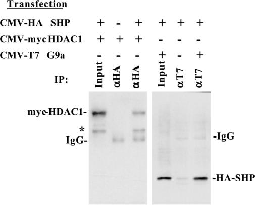
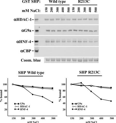
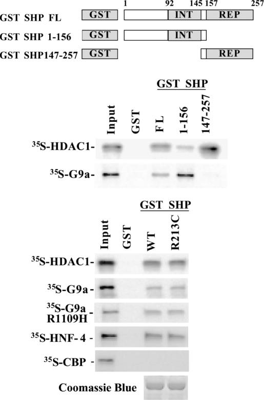
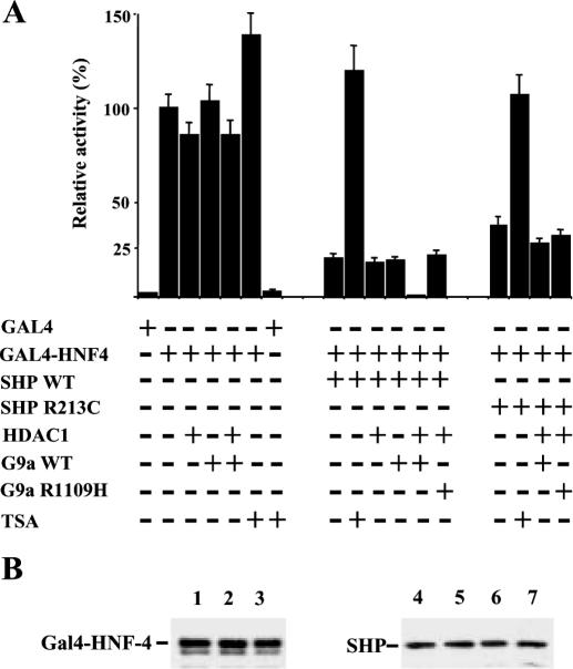
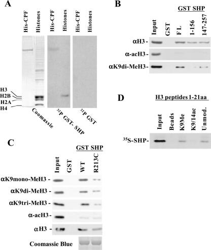
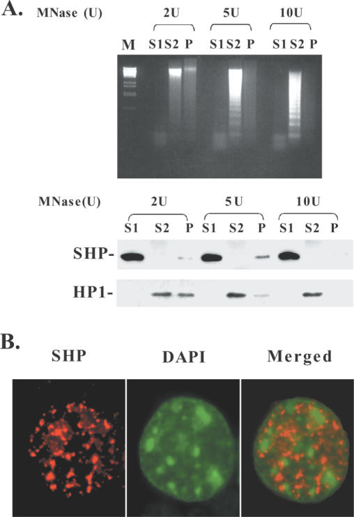
Site-specific modification of nucleosomal histones plays a central role in the formation of transcriptionally active and inactive chromatin structures. These modifications may serve as specific recognition motifs for chromatin proteins, which act as a signal for the adoption of the appropriate regulatory responses. Here, we show that the orphan nuclear receptor SHP (small heterodimer partner), a coregulator that inhibits the activity of several nuclear receptors, can associate with unmodified and lysine 9-methylated histone-3, but not with the acetylated protein. The naturally occurring SHP mutant (R213C), which exhibits decreased transrepression potential, interacts less avidly with K9-methylated histone 3. We demonstrate that SHP can functionally interact with histone deacetylase-1 and the G9a methyltransferase and that it is localized exclusively in nuclease-sensitive euchromatin. The results point to the involvement of a multistep mechanism in SHP-dependent transcriptional repression, which includes histone deacetylation, followed by H3-K9 methylation and stable association of SHP itself with chromatin.


Figures

Figure 1
Figure 1
Physical interaction of SHP with HDAC-1 and G9a in vivo. Nuclear extracts of Cos-1 cells transfected with the indicated expression vectors were immunoprecipitated with either αHA or αT7 antibodies. The immunoprecipitated materials were then analyzed in western blots with α-Myc antibody to detect myc-tagged HDAC-1 (left panel), or with αHA antibody to detect HA-tagged SHP proteins (right panel). The asterisk at left depicts an unidentified band, which probably corresponds to HDAC-1 degradation product.
Figure 2
Figure 2
SHP interacts with HDAC-1 and G9a with high affinity. GST pull-down experiments were performed with bacterially expressed GST-SHP fusion proteins and HepG2 nuclear extracts. The binding and wash buffers contained the indicated NaCl concentrations. Equal GST-fusion protein absorption to the beads was verified by Coomassie blue staining of a gel from a parallel experiment (bottom panel). The interacting proteins were identified by western blot analysis using the indicated antibodies. Serial exposures of enhanced chemiluminescence images were quantitated by the NIH-Image 1.63 software or by a Fujifilm LAS-1000 Luminescent Image analyzer and plotted as a percentage of the values obtained using buffers containing 150 mM NaCl.
Figure 3
Figure 3
Direct interaction of SHP with HDAC-1 and G9a in vitro. In vitro GST pull-down experiments were performed with 35S-labeled in vitro translated HDAC-1 or with G9a and bacterially expressed GST-SHP fusion proteins containing the indicated regions or the R213C mutation of the protein. INT depicts the receptor interaction domain of SHP (92–145 amino acids), while REP corresponds to the autonomous repression domain of SHP (157–257 amino acids). Equal coupling of wild-type and the R213C mutant GST-SHP proteins were verified by Coomassie blue staining.
Figure 4
Figure 4
Functional roles of HDAC-1 and G9a in SHP-mediated transcriptional repression. (A) Cos-1 cells were transfected with 0.5 μg 4xGal4-E1B-luc reporter together with 10 ng of SHP expression vectors and 100 ng of the other indicated expression vectors. Bars represent means ± SE of normalized luciferase activities, expressed as a percentage of the activity obtained by Gal4-HNF-4 alone (100%). Where indicated, the cells were treated with 0.1 μM trichostatin A (TSA) 12 h before harvest. (B) Whole cell extracts from Cos-1 cells were transfected with the above amounts of expression vectors and were analyzed in western blots using αHNF4 and αSHP antibodies, as indicated. The cells were transfected with vectors for Gal4-HNF-4 alone (lane 1), Gal-4-HNF-4 and SHPwt (lane 2), Gal-4-HNF-4, SHPwt, HDAC-1 and G9aWT (lane 3), SHPwt alone (lane 4), SHP R213C alone (lane 5), SHPwt, Gal4-HNF-4, HDAC-1 and G9aWT (lane 6), SHP R213C, Gal4-HNF-4, HDAC-1 and G9aWT (lane 7). The results show that under the conditions used, overexpression of the different proteins does not significantly alter the expression of Gal4-HNF4, SHPwt and SHP R213C.
Figure 5
Figure 5
Association of SHP with K9-methylated histone 3. (A) Core histones from HeLa cells were separated by SDS–PAGE, transferred to PVDF membranes and hybridized with 32P-GST as control (right panel), or 32P-GST-SHP (middle panel). Coomassie blue staining of the gel is shown at left. As a positive control for interaction, recombinant His-tagged CPF was run in parallel with histones (His-CPF lane). (B and C) An aliquot of 2 μg of the indicated GST-SHP fusion proteins linked to glutathione–sepharose beads were used to pull-down purified core histones. After washings, the retained material was subjected to western blot analysis with the indicated antibodies that detect total histone 3 (αH3), hyperacetylated histone 3 (α-acH3) and K9-mono, di or trimethylated histone 3 (αK9 mono-MeH3, αK9 di-MeH3, αK9 tri-MeH3). (D) Unmodified, K9-dimethylated (K9Me), or K9/K14-acetylated, biotin-conjugated histone 3 peptides (1–21 amino acids) were immobilized on streptavidin–agarose beads and used to pull-down 35S-labeled in vitro-translated SHP.
Figure 6
Figure 6
SHP is localized in euchromatic nuclear territories. (A) Caco-2 cell nuclei were treated with the indicated amounts of MNase and fractionated to obtain euchromatin-enriched (S1), heterochromatin-enriched (S2) and insoluble nuclear matrix (P) containing fractions. DNA from each fraction was purified and separated on 1.5% agarose gels (upper panel). Western blot analysis of the same fractions was performed with antibodies against SHP and HP1 (bottom panel). (B) In situ immunofluorescence analysis of Caco-2 cells was performed to detect SHP (left panel) and DAPI (middle panel). The merged image of the two stainings is shown at right.

References

    1. Turner B.M. (2000) Histone acetylation and an epigenetic code. BioEssays, 2, 836–845. - PubMed
    1. Jenuwein T. and Allis,C.D. (2001) Translating the histone code. Science, 93, 1074–1080. - PubMed
    1. Turner B.M. (2002) Cellular memory and the histone code. Cell, 111, 285–291. - PubMed
    1. Rea S., Eisenhaber,F., O'Carroll,D., Strahl,B.D., Sun,Z.W., Schmid,M., Opravil,S., Mechtler,K., Ponting,C.P., Allis,C.D. and Jenuwein,T. (2000) Regulation of chromatin structure by site-specific histone H3 methyltransferases. Nature, 406, 593–599. - PubMed
    1. Melcher M., Schmid,M., Aagaard,L., Selenko,P., Laible,G. and Jenuwein,T. (2000) Structure-function analysis of SUV39H1 reveals a dominant role in heterochromatin organization, chromosome segregation, and mitotic progression. Mol. Cell. Biol., 20, 3728–3741. - PMC - PubMed

Publication types

MeSH terms