Comparative Study
. 2004 Dec 10;95(12):1191-9.
doi: 10.1161/01.RES.0000150856.47324.5b. Epub 2004 Nov 18.

Cells expressing early cardiac markers reside in the bone marrow and are mobilized into the peripheral blood after myocardial infarction

Affiliations
Comparative Study

Cells expressing early cardiac markers reside in the bone marrow and are mobilized into the peripheral blood after myocardial infarction

Magda Kucia et al. Circ Res. .

Abstract

The concept that bone marrow (BM)-derived cells participate in cardiac regeneration remains highly controversial and the identity of the specific cell type(s) involved remains unknown. In this study, we report that the postnatal BM contains a mobile pool of cells that express early cardiac lineage markers (Nkx2.5/Csx, GATA-4, and MEF2C). These cells are present in significant amounts in BM harvested from young mice but their abundance decreases with age; in addition, the responsiveness of these cells to gradients of motomorphogens SDF-1, HGF, and LIF changes with age. FACS analysis, combined with analysis of early cardiac markers at the mRNA and protein levels, revealed that cells expressing these markers reside in the nonadherent, nonhematopoietic CXCR4+/Sca-1+/lin-/CD45- mononuclear cell (MNC) fraction in mice and in the CXCR4+/CD34+/AC133+/CD45- BMMNC fraction in humans. These cells are mobilized into the peripheral blood after myocardial infarction and chemoattracted to the infarcted myocardium in an SDF-1-CXCR4-, HGF-c-Met-, and LIF-LIF-R-dependent manner. To our knowledge, this is the first demonstration that the postnatal BM harbors a nonhematopoietic population of cells that express markers for cardiac differentiation. We propose that these potential cardiac progenitors may account for the myocardial regenerative effects of BM. The present findings provide a novel paradigm that could reconcile current controversies and a rationale for investigating the use of BM-derived cardiac progenitors for myocardial regeneration.


Figures

Figure 1
Figure 1
A, Detection of cardiac markers in BMMNCs that migrate to SDF-1, HGF, and LIF gradients. BM cells from 1-month-old mice were isolated from the lower transwell-chambers after chemo-taxis to SDF-1, HGF, and LIF (chemotactic isolation), and the expression of mRNA for cardiac markers (Nkx2.5/Csx and GATA-4) was compared by real-time RT-PCR between the same number of cells in the input (unpurified BM cells) and in the lower chamber. Data represent the average of three independent experiments (the BM of 10 mice was pooled for each experiment). Data are mean±SD. *P<0.00001 vs input cells. B, Immunocytochemical detection of protein expression for cardiac markers Nkx2.5/Csx and GATA-4 in input (unpurified) and SDF-1, HGF, and LIF chemoattracted BMMNCs from 1-month-old mice (the BM of 15 mice was pooled). BMMNCs migrating to an SDF-1 gradient exhibited greater abundance of cells positive for the cardiac markers. *P<0.05 vs input cells. C, Confocal microscopic images of BMMNCs positive for cardiac markers Nkx2.5/Csx (B, F, and J; red fluorescence), GATA-4 (A, E, and I; green fluorescence), and both (D, H, and L) in input (unpurified) BMMNCs (A through D) and BMMNCs chemoattracted to SDF-1 (E through H). I and J, Higher magnification images of a nucleus positive for both markers. Nuclei are identified by DAPI (C, D, G, H, K, and L; blue fluorescence). Scale bar=20 μm (A through H) or 10 μm (I through L).
Figure 2
Figure 2
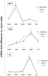
Expression of mRNA for cardiac markers in murine BMMNCs isolated by chemotaxis to SDF-1, HGF, and LIF at different ages. BMMNCs chemoattracted to SDF-1 (top panel), HGF (middle panel), and LIF (bottom panel) gradients were isolated from the lower transwell-chambers and the expression of mRNA for cardiac markers (Nkx2.5/Csx and GATA-4) was compared by real-time RT-PCR between the same number of cells in the input and in the lower chamber. All of the assays were performed in triplicate (the BM of 12 mice was pooled at each time-point). Data are mean±SD. *P<0.01 vs input.
Figure 3
Figure 3
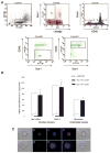
Expression of mRNA for cardiac and endothelial markers in murine Sca-1+/lin/CD45 cells (potential cardiac TCSCs). A, Dot-plot of murine BMMNCs (top left) and dot-plot of these cells from the lymphoid gate (R1) labeled for the expression of Sca-1 antigen and lineage negativity (top middle). Cells from the lymphoid gate that were Sca-1+/lin (R1, R2) were sorted by FACS for CD45 expression (top right). M1 shows CD45+ and M2 shows CD45 population of Sca-1+/lin cells. Dot-plot of Sca-1+/lin/CD45 is shown in bottom left panel (R5) and the dot-plot of Sca-1+/lin/CD45+ is shown in bottom right panel (R7). Four independent sorting experiments were performed (the BM of 12 mice was pooled for each sort). Representative sorts are shown. B, Real-time RT-PCR for the expression of mRNA for cardiac and endothelial markers. Markers were compared between the same number of sorted Sca-1+/lin/CD45+ and Sca-1+/lin/CD45 cells. Data represent the average of four independent experiments (the BM of 12 mice was pooled for each experiment). Data are mean±SD. *P<0.00001 vs unpurified BMMNCs. C, Representative images of BM-derived GATA-4 −positive Sca-1+/lin/CD45 cell (a through e, top panels) and GATA-4 −negative Sca-1+/lin/CD45+ cell (f through j, bottom panels) sorted by FACS. a and f, Phase-contrast image of cells; b and g, GATA-4 −positive (b, red fluorescence) and −negative (g) nuclei; c and h, Nuclei identified by DAPI (blue fluorescence); d and i, Overlay of b and c, and g and h, respectively; e and j, Overlay of the respective immunofluorescent and phase-contrast images. Scale bar=5 μm (all panels)
Figure 4
Figure 4
CD34+ and AC133+ human BMMNCs are highly enriched with mRNA for cardiac and endothelial markers. Human unpurified BMMNCs, adherent cell-depleted BMMNCs, and BMMNCs sorted by FACS for CD34− and AC133-positivity were compared for the expression of mRNA for early cardiac (Nkx2.5/Csx, GATA-4, and MEF2C) and endothelial (VE-cadherin, VEGFR3, and vWF) markers. mRNA was harvested separately from BMMNCs of 3 donors; for each donor, the assays were performed in triplicate. Data are mean±SD. *P<0.00001 vs control.
Figure 5
Figure 5
Changes in the expression of cardiac and endothelial markers in PB of mice after MI. PBMNCs were harvested from mice that underwent myocardial infarction or a sham operation (control) 6, 24, 48, and 96 hours earlier. Expression of mRNA for early cardiac (Nkx2.5/Csx, GATA-4, and MEF2C) and endothelial (VE-cadherin) markers in the same number cells was quantitated by real-time RT-PCR and compared between groups. All of the assays were performed in triplicate (PBMNCs from 8 to 10 mice per group were pooled at each time-point). Data are mean±SD. *P<0.01 vs control.
Figure 6
Figure 6
Myocardial expression of SDF-1, LIF, VEGF, and HGF at 6, 24, 48, and 96 hours after MI in mice. Expression of SDF-1, LIF, VEGF, and HGF mRNA in control (nonischemic zone, LV posterior wall) and infarcted (LV anterior wall) myocardium was quantitated by real-time RT-PCR. All of the assays were performed in triplicate (myocardial samples from 8 to 10 mice per group were examined at each time point). Data are mean±SD. *P<0.01 vs control.
Figure 7
Figure 7
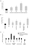
A, BMMNCs are chemoattracted to the supernatant from infarcted myocardial tissue in an SDF-1−, HGF−, and LIF-dependent manner. BMMNCs were added to the upper transwell chambers and allowed to migrate to the conditioned media (CM, supernatants) from control (LV posterior wall) and infarcted (LV anterior wall) myocardial tissue. BM of 5 mice was pooled, and the chemotactic assays were performed in triplicate. Data are mean±SD. *P<0.00001 vs control. B, Chemotaxis of BMMNCs to the conditioned media from control and infarcted myocardial tissue in the absence or presence of inhibitors to CXCR4 (T140), c-Met (K252a), and LIF-R (anti-gp190 Ab). BM of 5 mice was pooled, and the chemotactic assays were performed in triplicate. Data are mean±SD. *P<0.00001 vs CM from the infarcted myocardium. DMSO was added as control (solvent for K252a). C, Expression of mRNA for cardiac (Nkx2.5/Csx, GATA-4, and MEF2C), endothelial (VE-cadherin), and skeletal muscle (Myf5, MyoD, and myogenin) markers in control (unpurified) BMMNCs and BMMNCs that migrated to supernatants from infarcted myocardium. BM of 5 mice was pooled, and the chemotactic assays were performed in triplicate. Data are mean±SD. *P<0.0001 vs control.

References

    1. Orlic D, Kajstura J, Chimenti S, Jakoniuk I, Anderson SM, Li B, Pickel J, McKay R, Nadal-Ginard B, Bodine DM, Leri A, Anversa P. Bone marrow cells regenerate infarcted myocardium. Nature. 2001;410:701–705. - PubMed
    1. Orlic D, Kajstura J, Chimenti S, Limana F, Jakoniuk I, Quaini F, Nadal-Ginard B, Bodine DM, Leri A, Anversa P. Mobilized bone marrow cells repair the infarcted heart, improving function and survival. Proc Natl Acad Sci U S A. 2001;98:10344–10349. - PMC - PubMed
    1. Shake JG, Gruber PJ, Baumgartner WA, Senechal G, Meyers J, Redmond JM, Pittenger MF, Martin BJ. Mesenchymal stem cell implantation in a swine myocardial infarct model: engraftment and functional effects. Ann Thorac Surg. 2002;73:1919–1925. - PubMed
    1. Barbash IM, Chouraqui P, Baron J, Feinberg MS, Etzion S, Tessone A, Miller L, Guetta E, Zipori D, Kedes LH, Kloner RA, Leor J. Systemic delivery of bone marrow-derived mesenchymal stem cells to the infarcted myocardium: feasibility, cell migration, and body distribution. Circulation. 2003;108:863–868. - PubMed
    1. Strauer BE, Brehm M, Zeus T, Kostering M, Hernandez A, Sorg RV, Kogler G, Wernet P. Repair of infarcted myocardium by autologous intracoronary mononuclear bone marrow cell transplantation in humans. Circulation. 2002;106:1913–1918. - PubMed

Publication types

MeSH terms

Substances