. 2005 Jan;76(1):91-9.
doi: 10.1086/427266. Epub 2004 Nov 18.

Association between maternal age and meiotic recombination for trisomy 21

Affiliations

Association between maternal age and meiotic recombination for trisomy 21

Neil E Lamb et al. Am J Hum Genet. 2005 Jan.

Abstract

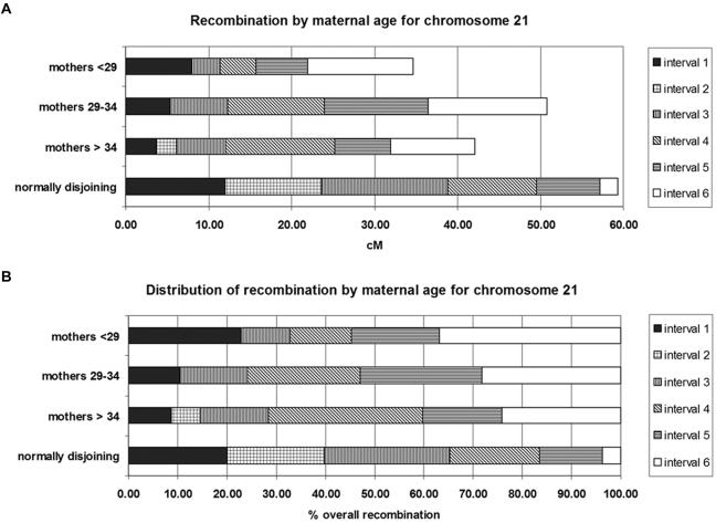
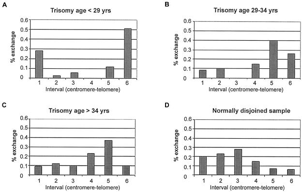
Altered genetic recombination has been identified as the first molecular correlate of chromosome nondisjunction in both humans and model organisms. Little evidence has emerged to link maternal age--long recognized as the primary risk factor for nondisjunction--with altered recombination, although some studies have provided hints of such a relationship. To determine whether an association does exist, chromosome 21 recombination patterns were examined in 400 trisomy 21 cases of maternal meiosis I origin, grouped by maternal age. These recombination patterns were used to predict the chromosome 21 exchange patterns established during meiosis I. There was no statistically significant association between age and overall rate of exchange. The placement of meiotic exchange, however, differed significantly among the age groups. Susceptible patterns (pericentromeric and telomeric exchanges) accounted for 34% of all exchanges among the youngest class of women but only 10% of those among the oldest class. The pattern of exchanges among the oldest age group mimicked the pattern observed among normally disjoining chromosomes 21. These results suggest that the greatest risk factor for nondisjunction among younger women is the presence of a susceptible exchange pattern. We hypothesize that environmental and age-related insults accumulate in the ovary as a woman ages, leading to malsegregation of oocytes with stable exchange patterns. It is this risk, due to recombination-independent factors, that would be most influenced by increasing age, leading to the observed maternal age effect.


Figures

Figure  1
Figure 1
Comparison of 21q interval boundaries between trisomic and normally disjoining samples. Interval distances for chromosome 21 markers are taken from the Ensembl Genome Browser (see Ensembl Web site), in which the “0 bp” position is at the telomere of the p arm and the most centromeric contig for the q arm begins at 13.3 Mb.
Figure  2
Figure 2
Recombination-based genetic maps of 21q for trisomic and normally disjoining samples. Genetic maps were created either directly on the Marshfield Web site (normally disjoining sample) or by use of the program NDJMap followed by application of the Kosambi map function to convert recombination fractions into map distances (trisomic sample). a, Overall map lengths divided into the six chromosome 21 intervals. b, Relative contribution of each interval to the entire map length.
Figure  3
Figure 3
Comparison of single-exchange events along chromosome 21q. The percentage of single exchanges in each chromosome interval is based on predictions from recombination data. The panels show the results of maternally inherited chromosomes that have undergone MI nondisjunction in women aged <29 years (A), 29–34 years (B), and >34 years (C) and chromosomes that segregated normally (D).
Figure  4
Figure 4
Examination of the location of the most-proximal meiotic exchange events along chromosome 21q. The location of the most proximal exchange was determined for each population, to examine the distance between the centromere and the closest meiotic exchange. The panels show the distribution for maternally inherited chromosomes that have undergone MI nondisjunction in women aged <29 years (A), 29–34 years (B), and >34 years (C) and for chromosomes that segregated normally (D).

References

Electronic-Database Information

    1. Ensembl, http://www.ensembl.org/ (for marker distances)
    1. Marshfield, http://research.marshfieldclinic.org/genetics/ (for genotype database)

References

    1. Broman K, Murray J, Sheffield V, White R, Weber J (1998) Comprehensive human genetic maps: individual and sex-specific variation in recombination. Am J Hum Genet 63:861–869 - PMC - PubMed
    1. Bugge M, Collins A, Petersen MB, Fisher J, Brandt C, Hertz JM, Tranebjaerg L, de Lozier-Blanchet C, Nicolaides P, Brondum-Nielsen K, Morton N, Mikkelsen M (1998) Non-disjunction of chromosome 18. Hum Mol Genet 7:661–66910.1093/hmg/7.4.661 - DOI - PubMed
    1. Feingold E, Brown AS, Sherman SL (2000) Multipoint estimation of genetic maps for human trisomies with one parent or other partial data. Am J Hum Genet 66:958–968 - PMC - PubMed
    1. Hassold T, Chiu D (1985) Maternal age–specific rates of numerical chromosome abnormalities with special reference to trisomy. Hum Genet 70:11–1710.1007/BF00389450 - DOI - PubMed
    1. Hassold T, Hunt P (2001) To err (meiotically) is human: the genesis of human aneuploidy. Nat Rev Genet 2:280–29110.1038/35066065 - DOI - PubMed

Publication types

Substances