

Lumbar incisional hernias: diagnostic and management dilemma
- PMID: 15554289
- PMCID: PMC3016827
Lumbar incisional hernias: diagnostic and management dilemma
Abstract
Introduction: Lumbar hernias occur infrequently and can be congenital, primary (inferior or Petit type, and superior or Grynfeltt type), posttraumatic, or incisional. They are bounded by the 12th rib, the iliac crest, the erector spinae, and the external oblique muscle. Most postoperative incisional hernias occur in nephrectomy or aortic aneurysm repair incisions.
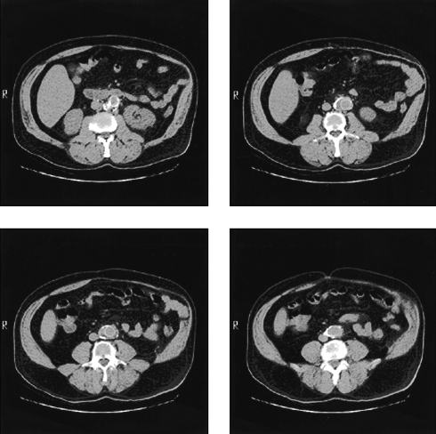
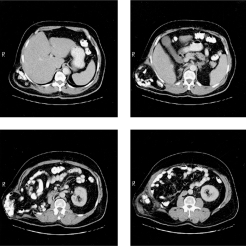
Case report: We present 2 patients who had undergone flank incisions and subsequently developed significant bulging of that area. The first patient had an atrophy of the abdominal wall musculature while the other had a large lumbar incisional hernia that was repaired laparoscopically.
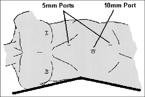
Discussion: Lumbar incisional hernias are often diffuse with fascial defects that are usually hard to appreciate. Computed tomography scan is the diagnostic modality of choice and allows differentiating them from abdominal wall musculature denervation atrophy complicating flank incisions. Repairing these hernias is difficult due to the surrounding structures. Principles of laparoscopic repair include lateral decubitus positioning with table flexed, adhesiolysis, and reduction of hernia contents, securing ePTFE mesh with spiral tacks and transfascial sutures to an intercostal space superiorly, iliac crest periosteum inferiorly, and rectus muscle anteriorly. Posteriorly, the mesh is secured to psoas major fascia with intracorporeal sutures to avoid nerve injury.
Conclusion: Lumbar incisional hernia must be differentiated from muscle atrophy with no fascial defect. The laparoscopic approach provides an attractive option for this often challenging problem.
Figures
References
-
- Barden BE, Maull KI. Traumatic lumbar hernia. South Med J. 2000;93(11):1067–1069 - PubMed
-
- Balkan M, Kozak O, Gulec B, Tasar M, Pekcan M. Traumatic lumbar hernia due to seat belt injury: case report. J Trauma. 1999;47(1):154–155 - PubMed
-
- Sarela Al, Mavanur AA, Bhaskar A, et al. Post traumatic lumbar hernia. J Postgrad Med. 1996;42(3):78–80 - PubMed
-
- Burick AJ, Parascandola SA. Laparoscopic repair of a traumatic lumbar hernia: a case report. J Laparoendosc Surg. 1996; 6(4):259–262 - PubMed
-
- Stevens KJ, Banuls M. Iliolumbar hernia following bone grafting. Eur Spine J. 1994;3(2):118–119 - PubMed
Publication types
MeSH terms
Substances
LinkOut - more resources
Full Text Sources