

T cell receptor induced intracellular redistribution of type I protein kinase A
- PMID: 15554923
- PMCID: PMC1782591
- DOI: 10.1111/j.1365-2567.2004.01992.x
T cell receptor induced intracellular redistribution of type I protein kinase A
Abstract
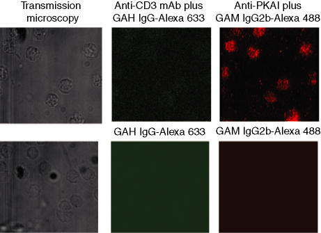
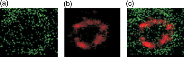
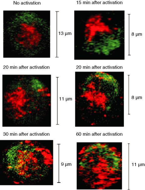
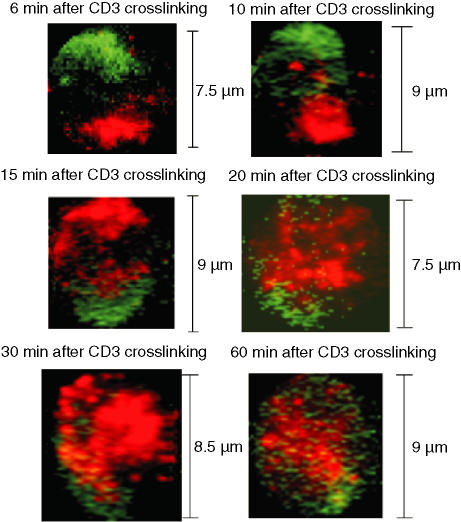
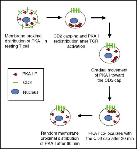
The productive activation of CD4(+) T lymphocytes, leading to proliferation and cytokine secretion, requires precise temporal regulation of intracellular cyclic AMP concentrations. The major effector molecule activated by cyclic AMP in mammalian cells is the cyclic AMP-dependent protein kinase A (PKA). The type I PKA isozyme mediates the inhibitory effects of cyclic AMP on T-cell activation. Using laser scanning confocal microscopy, we demonstrated that the regulation of PKA type I activity involves spatial redistribution of PKA type I molecules following T-cell receptor (TCR) stimulation. In resting T cells, PKA type I was located in membrane proximal regions and distributed equally across the cell. Shortly after antigen engagement, T cells and antigen-presenting cells formed an area of intense contact, known as the immunological synapse. TCR concentrated at the synapse, whereas PKA type I molecules redistributed to the opposite cell pole within 10 min after T-cell stimulation. Type I PKA redistribution was solely dependent on TCR signalling, because we observed the same temporal and spatial distribution after antibody-mediated cross-linking of the TCR-associated CD3 complex. Segregation of TCR and PKA type I molecules was maintained for at least 20 min. Thirty minutes after stimulation, PKA type I partially colocalized with the TCR. After 60 min, PKA type I distribution again approached the resting state. Considering that initial TCR signals lead to increases in intracellular cyclic AMP, PKA type I molecules may be targeted towards localized cyclic AMP accumulations or transported away from these areas, depending on the requirements of the cellular response.
Figures
Similar articles
-
Early events of human T lymphocyte activation are associated with type I protein kinase A activity.J Clin Invest. 1993 Nov;92(5):2207-14. doi: 10.1172/JCI116823. J Clin Invest. 1993. PMID: 8227335 Free PMC article.
-
Activation of type I protein kinase A during receptor-mediated human T lymphocyte activation.J Immunol. 1996 Jan 15;156(2):497-506. J Immunol. 1996. PMID: 8543799
-
[Inhibitory effects of HIV-1 gp41 fusion peptide on CD3 antibody activated regulatory T cells].Xi Bao Yu Fen Zi Mian Yi Xue Za Zhi. 2012 Nov;28(11):1121-5. Xi Bao Yu Fen Zi Mian Yi Xue Za Zhi. 2012. PMID: 23127396 Chinese.
-
Cyclic AMP-mediated immune regulation--overview of mechanisms of action in T cells.Cell Signal. 2011 Jun;23(6):1009-16. doi: 10.1016/j.cellsig.2010.11.018. Epub 2010 Dec 2. Cell Signal. 2011. PMID: 21130867 Review.
-
The molecular machinery for cAMP-dependent immunomodulation in T-cells.Biochem Soc Trans. 2006 Aug;34(Pt 4):476-9. doi: 10.1042/BST0340476. Biochem Soc Trans. 2006. PMID: 16856837 Review.
Cited by
-
Altered dynamics of Kv1.3 channel compartmentalization in the immunological synapse in systemic lupus erythematosus.J Immunol. 2007 Jul 1;179(1):346-56. doi: 10.4049/jimmunol.179.1.346. J Immunol. 2007. PMID: 17579055 Free PMC article.
-
The Ca(2+)-activated K(+) channel KCa3.1 compartmentalizes in the immunological synapse of human T lymphocytes.Am J Physiol Cell Physiol. 2007 Apr;292(4):C1431-9. doi: 10.1152/ajpcell.00376.2006. Epub 2006 Dec 6. Am J Physiol Cell Physiol. 2007. PMID: 17151145 Free PMC article.
-
Role of Phosphodiesterase 7 (PDE7) in T Cell Activity. Effects of Selective PDE7 Inhibitors and Dual PDE4/7 Inhibitors on T Cell Functions.Int J Mol Sci. 2020 Aug 25;21(17):6118. doi: 10.3390/ijms21176118. Int J Mol Sci. 2020. PMID: 32854348 Free PMC article. Review.
-
Compartmentalized Cyclic AMP Production by the Bordetella pertussis and Bacillus anthracis Adenylate Cyclase Toxins Differentially Affects the Immune Synapse in T Lymphocytes.Front Immunol. 2018 May 1;9:919. doi: 10.3389/fimmu.2018.00919. eCollection 2018. Front Immunol. 2018. PMID: 29765373 Free PMC article.
-
Trafficking of intermediate (KCa3.1) and small (KCa2.x) conductance, Ca(2+)-activated K(+) channels: a novel target for medicinal chemistry efforts?ChemMedChem. 2012 Oct;7(10):1741-55. doi: 10.1002/cmdc.201200226. Epub 2012 Aug 7. ChemMedChem. 2012. PMID: 22887933 Free PMC article. Review.
References
-
- Walsh DA, Perkins JP, Krebs EG. An adenosine 3′,5′-monophosphate-dependent protein kinase from rabbit skeletal muscle. J Biol Chem. 1968;243:3763–5. - PubMed
-
- Walsh DA, Van Patten SM. Multiple pathway signal transduction by the cAMP-dependent protein kinase. Faseb J. 1994;8:1227–36. - PubMed
-
- Corbin JD, Sugden PH, West L, Flockhart DA, Lincoln TM, McCarthy D. Studies on the properties and mode of action of the purified regulatory subunit of bovine heart adenosine 3′,5′-monophosphate-dependent protein kinase. J Biol Chem. 1978;253:3997–4003. - PubMed
-
- Doskeland SO, Ogreid D. Binding proteins for cyclic AMP in mammalian tissues. Int J Biochem. 1981;13:1–19. - PubMed
-
- Doskeland SO, Maronde E, Gjertsen BT. The genetic subtypes of cAMP-dependent protein kinase – functionally different or redundant? Biochim Biophys Acta. 1993;1178:249–58. - PubMed
Publication types
MeSH terms
Substances
Grants and funding
LinkOut - more resources
Full Text Sources
Molecular Biology Databases
Research Materials