. 2004 Dec;113(4):524-33.
doi: 10.1111/j.1365-2567.2004.01990.x.

Chemokine receptor expression in the human ectocervix: implications for infection by the human immunodeficiency virus-type I

Affiliations

Chemokine receptor expression in the human ectocervix: implications for infection by the human immunodeficiency virus-type I

Grant R Yeaman et al. Immunology. 2004 Dec.

Abstract

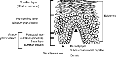
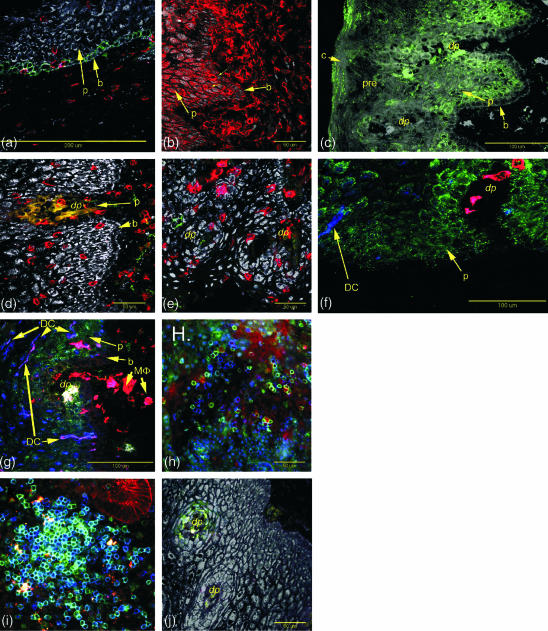
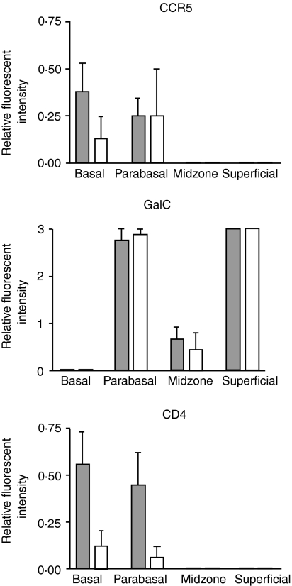
Human immunodeficiency virus-type 1 (HIV-1) is a sexually transmitted pathogen that can infect cells in the female reproductive tract (FRT). The mechanism of viral transmission within the FRT and the mode of viral spread to the periphery are not well understood. To characterize the frequency of potential targets of HIV infection within the FRT, we performed a systematic study of the expression of HIV receptors (CD4, galactosyl ceramide (GalCer)) and coreceptors (CXCR4 and CCR5) on epithelial cells and leucocytes from the ectocervix. The ectocervix is a likely first site of contact with HIV-1 following heterosexual transmission, and expression of these receptors is likely to correlate with susceptibility to viral infection. We obtained ectocervical tissue specimens from women undergoing hysterectomy, and compared expression of these receptors among patients who were classified as being in the proliferative or secretory phases of their menstrual cycle at the time of hysterectomy, as well as from postmenopausal tissues. Epithelial cells from tissues at early and mid-proliferative stages of the menstrual cycle express CD4, although by late proliferative and secretory phases, CD4 expression was absent or weak. In contrast, GalCer expression was uniform in all stages of the menstrual cycle. CXCR4 expression was not detected on ectocervical epithelial cells and positive staining was only evident on individual leucocytes. In contrast, CCR5 expression was detected on ectocervical epithelial cells from tissues at all stages of the menstrual cycle. Overall, our results suggest that HIV infection of cells in the ectocervix could most likely occur through GalCer and CCR5. These findings are important to define potential targets of HIV-1 infection within the FRT, and for the future design of approaches to reduce the susceptibility of women to infection by HIV-1.


Figures

Figure 1
Figure 1
The structure of non-keratinizing squamous epithelium in the ectocervix. The squamous mucosa of ectocervical squamous epithelium is divided into the indicated zones. The basal layer of cells (stratum basalis) together with the parabasal cells (stratum spinosum) are where cell division occurs and are collectively termed the stratum germinativum. The parabasal cells differentiate into the cells of the precornified layer or stratum granulosum. Further differentiation occurs forming the cornified layer (stratum corneum); here, the cells take on the characteristic cobblestone appearance of this epithelium. Beneath the basal lamina is the submucosal stroma that contains stromal cells and leucocytes, which traffic in from and out to the periphery. In certain areas, the underlying submucosal stroma exhibits finger like projections into the squamous mucosa known as submucosal stromal papillae that markedly reduce the distance between the submucosal stroma and the luminal surface.
Figure 2
Figure 2
CCR5 (green) is apparent on basal epithelial cells from some patients. CD3+ cells (red) in the same section do not express CCR5. This section was stained by the indirect method. CD4+ (red cells) cells are located in the stromal layer and squamous epithelium of the ectocervix (thin arrows). The basal (b) and parabasal (p) epithelial cells also show low levels of CD4 expression. GalCer (green) is expressed on parabasal epithelial cells (p) and in the surface regions of the cornified layer (c) but not on the basal layer (b) or precornified layer (pre). CD8+ T cells were present in both the submucosal stroma and the squamous epithelium. In comparison to CD4+ cells, they penetrated further into the basal and mid- zones. No CCR5 expression was observed on these cells by direct staining. In contrast to CD163 (f), CD14 expression is found on both stroma and squamous epithelium macrophages. CCR5+ (green) cells are found in association with the margins of the submucosal stromal papillae (dp). CD163 cells (red) are evident in the submucosal stroma and submucosal stroma papillae but are largely absent from the squamous epithelium. CD1a positive dendritic cells (DC) are present in the squamous epithelium (blue). Concentrations of HLA-DR+ cells (red) are associated with the submucosal stromal papillae some of which express GalCer (yellow cells in the papillae). CD1a+ HLA-DR+ dendritic cells (DC, purple colour) were present in the adjacent squamous epithelium. CCR5+ (green) and T cells (CD3, red) are readily detectable in the submucosal stroma using fresh unfixed vibratome sections of ectocervical tissue. Proliferative phase endometrium from a patient exhibiting intense staining for CCR5 (green) on T cells (CD3, blue) appearing turquoise. CCR5 positive cells (green) in submucosal stromal papillae (dp).
Figure 3
Figure 3
Expression of CCR5, GalCer and CD4 in the different epithelial zones during the proliferative and secretory phases of the menstrual cycle. Proliferative phase tissues, filled bars (n = 10), secretory phase tissues, open bars (n = 8). Error bars are 1 sd. The data shown for CCR5 were determined using the indirect staining protocol.

References

    1. Yeaman GR, Howell AL, Weldon S, et al. HIV receptor and co-receptor expression on human uterine epithelial cells: Regulation during the menstrual cycle and implications for HIV infection. Immunology. 2003;109:137–46. - PMC - PubMed
    1. Poppe W, Drijkoningen M, Ide P, Lauweryns J, Van Assche F. Lymphocytes and dendritic cells in the normal uterine cervix. An immunohistochemical study. Reprod Biol. 1998;81:277–82. - PubMed
    1. Roncalli M, Sideri M, Gie P, Servida E. Immunophenotypic analysis of the transformation zone of human cervix. Lab Invest. 1988;58:141–9. - PubMed
    1. Gupta P, Collins KB, Ratner D, Watkins S, Naus GJ, Landers DV, Patterson BK. Memory CD4+ T cells are the earliest detectable human immunodeficiency virus type 1 (HIV-1)-infected cells in the female genital mucosal tissue during HIV-1 transmission in an organ culture system. J Virol. 2002;76:9868–76. - PMC - PubMed
    1. Hu J, Gardner MD, Miller CJ. Simian immunodeficiency virus rapidly penetrates the cervicovaginal mucosa after intravaginal inoculation and infects intraepithelial dendritic cells. J Virol. 2000;74:6087–95. - PMC - PubMed

Publication types