. 2004 Dec;54(5):1199-211.
doi: 10.1111/j.1365-2958.2004.04345.x.

Energetic components VirD4, VirB11 and VirB4 mediate early DNA transfer reactions required for bacterial type IV secretion

Affiliations

Energetic components VirD4, VirB11 and VirB4 mediate early DNA transfer reactions required for bacterial type IV secretion

Krishnamohan Atmakuri et al. Mol Microbiol. 2004 Dec.

Abstract

Bacteria use type IV secretion systems (T4SS) to translocate DNA (T-DNA) and protein substrates across the cell envelope. By transfer DNA immunoprecipitation (TrIP), we recently showed that T-DNA translocates through the Agrobacterium tumefaciens VirB/D4 T4SS by forming close contacts sequentially with the VirD4 receptor, VirB11 ATPase, the inner membrane subunits VirB6 and VirB8 and, finally, VirB2 pilin and VirB9. Here, by TrIP, we show that nucleoside triphosphate binding site (Walker A motif) mutations do not disrupt VirD4 substrate binding or transfer to VirB11, suggesting that these early reactions proceed independently of ATP binding or hydrolysis. In contrast, VirD4, VirB11 and VirB4 Walker A mutations each arrest substrate transfer to VirB6 and VirB8, suggesting that these subunits energize this transfer reaction by an ATP-dependent mechanism. By co-immunoprecipitation, we supply evidence for VirD4 interactions with VirB4 and VirB11 independently of other T4SS subunits or intact Walker A motifs, and with the bitopic inner membrane subunit VirB10. We reconstituted substrate transfer from VirD4 to VirB11 and to VirB6 and VirB8 by co-synthesis of previously identified 'core' components of the VirB/D4 T4SS. Our findings define genetic requirements for DNA substrate binding and the early transfer reactions of a bacterial type IV translocation pathway.


Figures
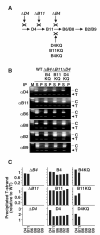
Fig. 1
Fig. 1
Effects of Walker A mutations on substrate transfer through the VirB/D4 T4SS. A. Effects of null mutations and Walker A mutations of the three energetic subunits on substrate transfer through the postulated translocation pathway. B. T-strand interactions with T4SS subunits from strains producing Walker A mutant proteins as shown by TrIP. Strains: wild type (WT), A348; ΔB4(B4KQ), PC1004(pKA102) producing VirB4K439Q; ΔB11(B11KQ), PC1011(pSR40) producing VirB11K175Q; ΔD4(D4KQ), Mx355(pKA101) producing VirD4K152Q. Strains were treated in vivo with formaldehyde before lysis. Antibodies listed on the left were used to immunoprecipitate (IP) the cognate Vir protein and associated DNA substrate; S, supernatant after precipitation; P, precipitate. Samples were assayed for T-strand (T) and the pTi control fragment (C) by PCR amplification and agarose gel electrophoresis. C. Results of quantitative TrIP (QTrIP) assay. Bars in the histogram represent the amount of T-strand recovered with antibodies to a given T4SS subunit (listed on the x-axis) from a mutant strain (identified above each histogram panel) relative to that recovered with the same antibody from the isogenic WT strain (normalized to 1.0). Strains: ΔB4, PC1004; ΔB4(B4), PC1004(pKA93) producing VirB4; ΔB4(B4KQ), PC1004(pKA102) producing VirB4K439Q; ΔB11, PC1011; ΔB11(B11), PC1011(pSR45) producing VirB11; ΔB11(B11KQ), PC1011(pSR40) producing VirB11K175Q; ΔD4, Mx355; ΔD4(D4), Mx355(pKA42) producing VirD4; ΔD4(D4KQ), Mx355(pKA101) producing VirD4K152Q. Note that the VirD4 antibodies precipitated T-strand from Mx355(pKA42) or Mx355(pKA101) at levels approximately three- or twofold higher than from the WT strain. These strains overproduce the VirD4 proteins as a result of gene expression from a pBBR1-based replicon, whose copy number is estimated to be several-fold higher than the native pTi plasmid (Kovach et al., 1994).
Fig. 2
Fig. 2
Co-immunoprecipitation of complexes composed of VirD4 T4CP, VirB10, VirB4 and VirB11. A. Co-immunoprecipitation of complexes from wild-type cell extracts. Material immunoprecipitated (IP) with preimmune serum (Pre) or antibodies to the proteins listed (αD4, etc.) were analysed by Western blotting for the presence of Vir proteins listed on the right. (*) VirB10 undergoes proteolysis to an ≈40 kDa species; other unlabelled bands correspond to the cross-reactive heavy chain IgG. Total DDAO-solubilized membrane proteins (MP, right) from the WT strain show positions of the Vir proteins. Molecular mass markers (M) and sizes in kilodaltons (kDa) are shown on the left. B. Co-immunoprecipitation of VirD4 and VirB proteins with VirD4 antibodies from extracts of strains deleted of virD4D4), virB4B4), virB10B10) or virB11B11).
Fig. 3
Fig. 3
ATP-binding subunits interact independently of other T4SS subunits and energization. A. Co-immunoprecipitation of VirD4, VirB4 and VirB11 in the absence of other T4SS subunits. Material immunoprecipitated (IP) with antibodies to the proteins listed (αD4, etc.) were analysed by Western blotting for the presence of Vir proteins listed on the right. Unlabelled bands correspond to the cross-reactive heavy chain IgG. Total DDAO-solubilized membrane proteins (MP, right) from the WT strain show positions of the Vir proteins. Strains: ΔvirB (PC1000), producing B4 (pKA93); B11 (pSR45); B4 and B11 (pYJB61). Molecular mass markers (M) and sizes in kilodaltons (kDa) are shown on the left. B. Immunoprecipitation with VirD4 antibodies of proteins from strains producing combinations of native or Walker A mutants. Left blots: ΔvirB, PC1000 producing VirD4, and either B4KQ (VirB4K439Q from pKA102) or B11KQ (VirB11K175Q from pSR40). Right blots: ΔD4 ΔvirB (KA2001), producing D4KQ (VirD4K152Q from pKA101); D4KQ and B4 (pKA101, pTAD214); D4KQ and B11 (pKA101, pSR45); D4KQ and B4 and B11 (pKA101, pYJB61). C. Co-immunoprecipitation of VirB4 and VirB11 with antibodies to each protein in the absence of VirD4 ΔB4, PC1004; ΔB11, PC1011; ΔD4, Mx355; ΔD4 ΔvirB, KA2001, producing B4 and B11 (pYJB61).
Fig. 4
Fig. 4
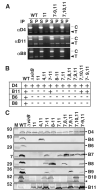
in vivo reconstitution of substrate transfer from VirD4 to VirB11. A. T-strand interactions with T4SS subunits from strains producing subsets of VirB proteins as shown by TrIP. Strains: WT, A348; 11, KA1001; 7–9, 11, KA1009; 7, 10, 11, KA1008. Strains were treated and samples were analysed as in Fig. 1. Precipitation of T-strand substrate (T) and the pTi control fragment (C) was detected by PCR amplification and agarose gel electrophoresis. B. Summary of strains analysed by TrIP for reconstitution of T-strand transfer from VirD4 to VirB11. KA10XX strains (see Experimental procedures) produce the VirB proteins listed at the top, as confirmed by Western blot analysis (Fig. 4C). (+) T-strand amplification product detected by agarose gel electrophoresis; (−) no detectable amplification product. C. VirB and VirD4 protein accumulation in A348 (WT), PC1000 (ΔvirB) and the KA10XX strains engineered to produce the VirB proteins listed vertically. Equivalent amounts of total membrane proteins from strains induced for vir gene expression to an OD600 = 0.5 in ABIM were analysed for Vir protein content by SDS-PAGE and Western blotting with antibodies to the VirB and VirD4 proteins listed on the right. (*) a VirB10 proteolytic product. Molecular mass markers (M) and sizes in kilodaltons (kDa) are shown on the left.
Fig. 5
Fig. 5
in vivo reconstitution of substrate transfer to VirB6 and VirB8. A. T-strand interactions with T4SS subunits from strains producing subsets of VirB proteins as shown by TrIP. Strains: WT, A348; 6–11, KA1010; 4, 6–11, KA1011; 4, 6–9, 11, KA1012; 4, 6, 7, 10, 11, KA1013; 4, 6–8, 10, 11, KA1014; 4, 6–8, 11; KA1015. B. Summary of strains analysed by TrIP for reconstitution of T-strand transfer from VirD4 to VirB6 and VirB8. C. VirB and VirD4 protein accumulation in strains producing the VirB proteins listed vertically. Samples were prepared and analysed as described in the legend to Fig. 4C. Immunoblots were developed with antibodies to the VirB and VirD4 proteins listed on the right. (*) a VirB10 proteolytic product. Molecular mass markers (M) and sizes in kilodaltons (kDa) are shown on the left.
Fig. 6
Fig. 6
Steps depicting early stages of DNA translocation through the A. tumefaciens VirB/D4 T4SS. VirD4 T4CP recruits the T-DNA (step I) and delivers substrate to VirB11 (step II). Next, two models are envisioned (step III). A ‘ping-pong’ model depicts VirB11 as a chaperone that unfolds the relaxase and retrotransfers the unfolded substrate back to VirD4. VirD4 then translocates the unfolded relaxase-T-strand complex across the inner membrane where it then accesses the VirB channel through a vestibule formed by a periplasmic loop of VirB6 (see Jakubowski et al., 2004). An alternative ‘shoot-and-pump’ model postulates that VirB11 interacts with other VirB proteins to transfer the relaxase component of the transfer intermediate across the inner membrane at the same time as VirD4 mediates transfer of the T-strand component (adapted from Llosa et al., 2002). For both models, the early transfer reactions (steps I and II) proceed independently of ATP utilization, whereas successive reactions depicted by the two models (step III) are regulated by ATP through the co-ordinated activities of VirD4, VirB11 and VirB4.

References

    1. Atmakuri K, Ding Z, Christie PJ. VirE2, a type IV secretion substrate, interacts with the VirD4 transfer protein at cell poles of Agrobacterium tumefaciens. Mol Microbiol. 2003;49:1699–1713. - PMC - PubMed
    1. Berger BR, Christie PJ. The Agrobacterium tumefaciens virB4 gene product is an essential virulence protein requiring an intact nucleoside triphosphate-binding domain. J Bacteriol. 1993;175:1723–1734. - PMC - PubMed
    1. Berger BR, Christie PJ. Genetic complementation analysis of the Agrobacterium tumefaciens virB operon: virB2 through virB11 are essential virulence genes. J Bacteriol. 1994;176:3646–3660. - PMC - PubMed
    1. Binns A, Beaupre C, Dale E. Inhibition of VirB-mediated transfer of diverse substrates from Agrobacterium tumefaciens by the IncQ plasmid RSF1010. J Bacteriol. 1995;177:4890–4899. - PMC - PubMed
    1. Cabezon E, Lanka E, de la Cruz F. Requirements for mobilization of plasmids RSF1010 and ColE1 by the IncW plasmid R388: trwB and RP4 traG are interchangeable. J Bacteriol. 1994;176:4455–4558. - PMC - PubMed

Publication types

MeSH terms