Comparative Study
. 2005 Feb;88(2):1311-21.
doi: 10.1529/biophysj.104.050203. Epub 2004 Nov 19.

A double-deletion method to quantifying incremental binding energies in proteins from experiment: example of a destabilizing hydrogen bonding pair

Affiliations
Comparative Study

A double-deletion method to quantifying incremental binding energies in proteins from experiment: example of a destabilizing hydrogen bonding pair

Luis A Campos et al. Biophys J. 2005 Feb.

Abstract

The contribution of a specific hydrogen bond in apoflavodoxin to protein stability is investigated by combining theory, experiment and simulation. Although hydrogen bonds are major determinants of protein structure and function, their contribution to protein stability is still unclear and widely debated. The best method so far devised to estimate the contribution of side-chain interactions to protein stability is double mutant cycle analysis, but the interaction energies so derived are not identical to incremental binding energies (the energies quantifying net contributions of two interacting groups to protein stability). Here we introduce double-deletion analysis of 'isolated' residue pairs as a means to precisely quantify incremental binding. The method is exemplified by studying a surface-exposed hydrogen bond in a model protein (Asp96/Asn128 in apoflavodoxin). Combined substitution of these residues by alanines slightly destabilizes the protein due to a decrease in hydrophobic surface burial. Subtraction of this effect, however, clearly indicates that the hydrogen-bonded groups in fact destabilize the native conformation. In addition, molecular dynamics simulations and classic double mutant cycle analysis explain quantitatively that, due to frustration, the hydrogen bond must form in the native structure because when the two groups get approximated upon folding their binding becomes favorable. We would like to remark that 1), this is the first time the contribution of a specific hydrogen bond to protein stability has been measured by experiment; and 2), more hydrogen bonds need to be analyzed to draw general conclusions on protein hydrogen bond energetics. To that end, the double-deletion method should be of help.


Figures

FIGURE 1
FIGURE 1
Energy inventory in double-mutant cycle and double-deletion analyses. The equations show the relationship between the incremental binding energy from the unfolded state (the contribution of any two groups to protein stability), the double-mutant cycle interaction energy, and the double-deletion energy. See Theory.
FIGURE 2
FIGURE 2
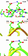
(A) Ball and stick representation of the Anabaena apoflavodoxin structure (1ftg) showing the hydrogen bonded residues D96 and N128. Hydrogen bonds in magenta. (B) Superposition of the apoflavodoxin from Anabaena and holo flavodoxin from Chondrus crispus (2fcr) showing the Anabaena hydrogen-bonded residues D96 and N128 and their structural equivalents, D100 and E132. The perfect conservation of the structure at the site of mutation in the Chondrus crispus protein, where the hydrogen bond is no longer possible, can be appreciated. (C) Superposition of the Anabaena apo and holo (1flv) flavodoxin structures showing the conservation of the hydrogen bond upon FMN cofactor binding.
FIGURE 3
FIGURE 3
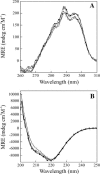
Near-UV (A) and far-UV (B) circular dichroism spectra of wild-type (solid circles), D96A (solid triangles), N128A (open circles), and D96A/N128A (open triangles) mutants. Spectra obtained at 25.0 ± 0.1°C in MOPS, 50 mM, pH 7.0.
FIGURE 4
FIGURE 4
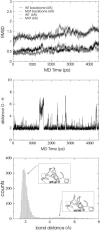
Molecular dynamics simulation of wild-type Anabaena apoflavodoxin and of the D96A/N128A double mutant. (Top) RMSD of the overall structures (upper traces) and of the atoms within a 6-Å radius of the carboxyl and carboxamide groups removed upon mutation (lower traces). The initial raising of the RMSD traces corresponds to the initial heating to 298 K of the reference minimized structures. (Middle) Evolution of the hydrogen bond H…O distance along the simulation. The shorter of the distances between the side-chain NH hydrogen of N128 and any of the side-chain O atoms of D96 is represented. Hydrogen bond breaking and reforming events are evidenced as peaks from the equilibrium distance baseline. (Bottom) Statistics of hydrogen bond distances during a 4.5-ns simulation of wild-type apoflavodoxin. Counts of distances sampled every picosecond are shown. The main peak represents the conformations that retain the hydrogen bond (see inset) whereas the flatter, wider peak represents conformations with a broken hydrogen bond. A 2.5 ± 0.1-Å cutoff has been used to calculate the free energy of hydrogen bond formation from the folded state.
FIGURE 5
FIGURE 5
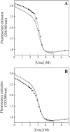
Urea denaturation curves of wild-type (solid circles) and D96A/N128A (open circles) apoflavodoxin double mutant (A) and of the D96A (solid circles) and N128A (open circles) single mutants (B). Data were recorded at 25.0 ± 0.1°C in MOPS, 50 mM, pH 7.0, with 0.5 M NaCl, and fitted to a two-state equation (Santoro and Bolen, 1988).
FIGURE 6
FIGURE 6
Scheme depicting the folding of a protein as divided into two steps. In the first one, with ΔGI, the protein gets folded to a virtual intermediate (essentially folded) where the i and j residues do not yet establish an interaction. Here, the solvations of the i and j residues are equivalent to those in the folded state of the 0j and i0 single mutants, and the interaction between them is considered close to zero. In the second step, with ΔGII, the two residues establish an interaction. The equations show the relationship between ΔGII, which represents the incremental binding energy from the virtual, folded, intermediate, and interaction energy, calculated from double-mutant cycle analysis.

References

    1. Ben-Tal, N., D. Sitkoff, I. A. Topol, A.-S. Yang, S. Buro, and B. Ong. 1997. Free energy of amide hydrogen bond formation in vacuum, in water, and in liquid alkane solution. J. Phys. Chem. B. 101:450–457.
    1. Blaber, M., J. D. Lindstrom, N. Gassner, J. Xu, D. W. Heinz, and B. W. Matthews. 1993. Energetic cost and structural consequences of burying a hydroxyl group within the core of a protein determined from Ala→Ser and Val→Thr substitutions in T4 lysozyme. Biochemistry. 32:11363–11373. - PubMed
    1. Branden, C., and J. Tooze. 1998. Introduction to Protein Structure. Garland Publishing, New York.
    1. Brooks, B. R., R. E. Bruccoleri, B. D. Olafson, D. J. Status, S. Swaminathan, and M. J. Karplus. 1983. CHARMM: a program for macromolecular energy minimization and dynamics calculations. J. Comput. Chem. 4:187–217.
    1. Carter, P. J., G. Winter, A. J. Wilkinson, and A. R. Fersht. 1984. The use of double mutants to detect structural changes in the active site of the tyrosyl-tRNA synthetase (Bacillus stearothermophilus). Cell. 38:835–840. - PubMed

Publication types

MeSH terms

LinkOut - more resources