. 2004 Nov 30;101(48):16801-6.
doi: 10.1073/pnas.0407647101. Epub 2004 Nov 19.

Reversible transdifferentiation of secretory epithelial cells into adipocytes in the mammary gland

Affiliations

Reversible transdifferentiation of secretory epithelial cells into adipocytes in the mammary gland

Manrico Morroni et al. Proc Natl Acad Sci U S A. .

Abstract

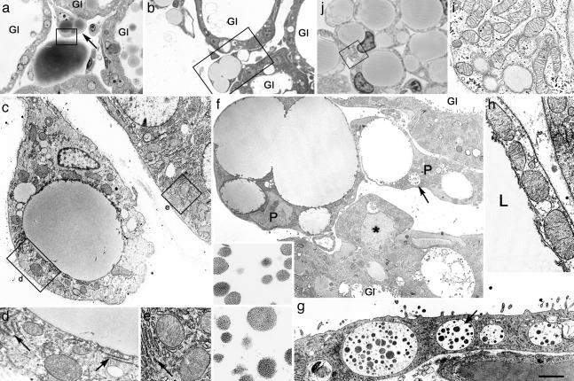
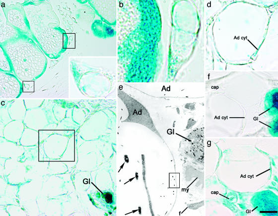
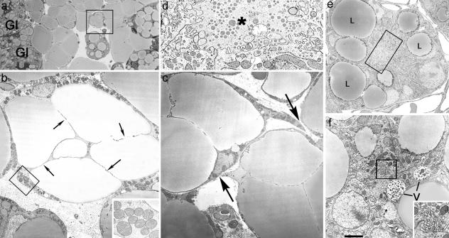
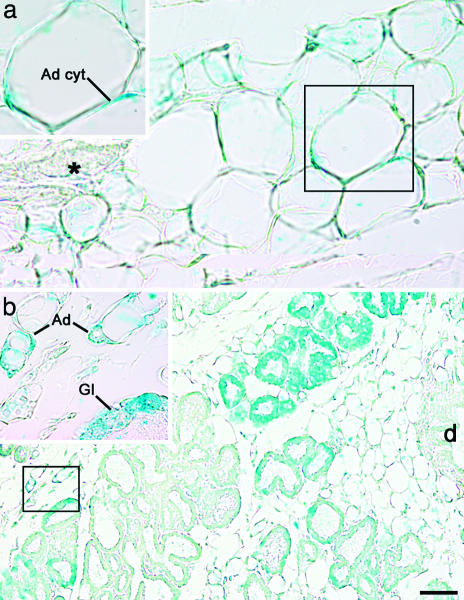
Mammalian breast adipose tissue is replaced by a milk-secreting gland during pregnancy; the reverse process takes place upon interruption of lactation. Morphological and bromodeoxyuridine studies provide indirect evidence that mouse mammary adipocytes transform into secretory epithelial cells during pregnancy and revert to adipocytes after lactation. By using the Cre-loxP recombination system we show that the mammary gland of whey acidic protein (WAP)-Cre/R26R mice, in which secretory epithelial cells express the lacZ gene during pregnancy, contains labeled adipocytes during involution. Conversely, adipocyte P2-Cre/R26R mice, in which adipocytes are labeled before pregnancy, contain labeled secretory epithelial cells during pregnancy. We conclude that reversible adipocyte-to-epithelium and epithelium-to-adipocyte transdifferentiation occurs in the mammary gland of adult mice during pregnancy and lactation.


Figures

Fig. 1.
Fig. 1.
During postlactation involution, adipocyte precursors among mammary gland alveoli show epithelial features and differ from typical white adipocyte precursors. (ah) Day 1 of postlactation involution. (a and b) Light microscopy of unilocular and multilocular adipocyte precursors among mammary gland alveoli (Gl). (c) Electron microscopy of the adipocyte precursor indicated by arrow in a; part of the alveolar epithelium is also visible. (d and e) Enlargements of the areas boxed in c, showing similarities of mitochondria and stacked rough endoplasmic reticulum (RER) (arrows) in the two cell types. (f) Electron microscopy of the area boxed in b: adipocyte precursors (P), one of which contains vacuoles (arrow) filled with milk protein secretory granules (enlarged in Inset) identical to those found in alveolar epithelium (compare with g). An alveolar epithelial cell protruding toward the interstitium (asterisk) is also visible. Gl, glands. (g) Electron microscopy of the alveolar epithelium with milk protein secretory granules (indicated by arrow and enlarged in Inset). (h) Electron microscopy of the area boxed in a: mitochondria of the mature unilocular adipocyte (at left) are large and similar to those of the adipocyte precursor (at right) and of mammary gland alveolar epithelium (compare with e). L, lipid droplet. (i and j) Developing mammary gland of a 12-h-old mouse. (i) The area boxed in j. Under the electron microscope, typical adipocyte precursors show the classic elongated mitochondria, which are distinctly different from those observed in mammary gland adipocyte precursors during postlactation adipogenesis (compare with d). [Scale bar: 10 (a), 12.5 (b), 1.2 (c), 0.8 (d, e, and i), 2.4 (f), 0.3 (Inset in f and g), 2 (g), 1 (h), and 7 (j) μm.]
Fig. 2.
Fig. 2.
Postlactation adipocytes express the gene reporter driven by the alveolar epithelial cell-specific WAP promoter. (af) Mammary gland of WAP-Cre/R26R mouse, β-gal histochemistry. (a and b) On day 1 of postlactation involution, epithelial alveolar cells of the gland as well as adipocyte precursors (boxed) stain for X-Gal. (a Inset) Enlargement of the lower of the two boxed areas in a. (b) Enlargement of the upper of the two boxed areas in a. (ce) On day 10 of postlactation involution, atrophic glands and most adipocytes are positive for X-Gal. (c) Gl, atrophic glands. (d) Enlargement of the area boxed in c. The cytoplasm of the adipocyte (Ad cyt) is X-Gal-positive. (e) By electron microscopy, typical X-Gal crystals are present only in adipocytes (Ad) and the epithelial component of the atrophic gland (Gl). Note the absence of crystals in other cell types. my, Myoepithelial cell; f, fibroblast. (e Inset) Enlargement of boxed area in e showing the typical crystals (arrows). (f) On day 9 of pregnancy, only the epithelial component of the mammary gland (Gl) is positive, whereas adipocytes (Ad cyt) and the other cell types [such as capillary (cap)] do not stain for X-Gal (negative control). (g) In a ROSA26 mouse (positive control) on day 1 of postlactation involution, all of the cell types found in the mammary gland were positive. Ad cyt, adipocyte cytoplasm; cap, capillary; Gl, gland. [Scale bar: 17 (a), 4 (a Inset), 2 (b), 9 (c), 4 (d, f, and g), 2 (e), and 0.4 (e Inset) μm.]
Fig. 3.
Fig. 3.
During pregnancy, adipocytes of the mammary gland exhibit epithelial features. Mouse mammary gland on day 18 of pregnancy. (a) Light microscopy. (bf) Electron microscopy. (a) Adipocytes near the newly formed alveoli (Gl) show lipid droplet compartmentalization. (b) Electron microscopy of the area squared in a. Adipocytes show a thickened peripheral cytoplasm containing unusually large mitochondria (enlarged in Inset) and thin cytoplasmic processes (arrows) that seem to subdivide the lipid droplet. (c) Transdifferentiating adipocytes also exhibit cytoplasmic projections (arrows), suggesting a tendency to become joined and to give rise to early alveolar structures (see df). (d) Enlargement of the boxed area in e, showing a small lumen (asterisk) with microvilli. (e) Early alveolar structure containing large lipid droplets (L). (f) Vacuoles (V) containing milk protein secretory granules are present in some early alveolar structures (see the lumen crowded with microvilli in the boxed area, which is enlarged in Inset). [Scale bar: 15 (a), 2.5 (b), 0.5 (b Inset), 2.5 (c), 0.5 (d), 2 (e), 1.2 (f), and 0.4 (f Inset) μm.]
Fig. 4.
Fig. 4.
During pregnancy, mammary alveolar epithelial cells express the gene reporter driven by the adipocyte-specific aP2 promoter. Mammary gland of aP2-Cre/R26R mouse, β-gal histochemistry. (a) In virgin mice only adipocytes stain for X-Gal. Note the absence of staining in the epithelial portion of the gland (asterisk). Ad cyt, adipocyte cytoplasm. (b) On day 18 of pregnancy, adipocytes (Ad) as well as many alveolar epithelial cells (Gl) are positive for X-Gal. d, Main duct. (Insets) Enlargements of the corresponding boxed areas. [Scale bar: 10 (a), 8 (Insets), and 30 (b) μm.]

References

    1. Hennighausen, L. & Robinson, G. W. (2001) Dev. Cell 1, 467–475. - PubMed
    1. Silberstein, G. B. (2001) Microsc. Res. Tech. 52, 155–162. - PubMed
    1. Tosh, D. & Slack, J. M. W. (2002) Nat. Rev. Mol. Cell Biol. 3, 187–194. - PubMed
    1. Soriano, P. (1999) Nat. Genet. 21, 70–71. - PubMed
    1. Wagner, K.-U., Wall, R. J., St-Onge, L., Gruss, P., Wynshaw-Boris, A., Garret, L., Li, M., Furth, P. A. & Hennighausen, L. (1997) Nucleic Acids Res. 25, 4323–4330. - PMC - PubMed

Publication types