. 2004 Dec;136(4):4037-47.
doi: 10.1104/pp.104.052357. Epub 2004 Nov 19.

Polyphyletic origin of pyrrolizidine alkaloids within the Asteraceae. Evidence from differential tissue expression of homospermidine synthase

Affiliations

Polyphyletic origin of pyrrolizidine alkaloids within the Asteraceae. Evidence from differential tissue expression of homospermidine synthase

Sven Anke et al. Plant Physiol. 2004 Dec.

Abstract

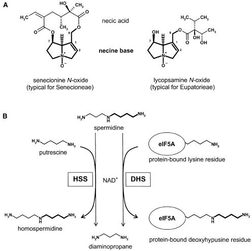
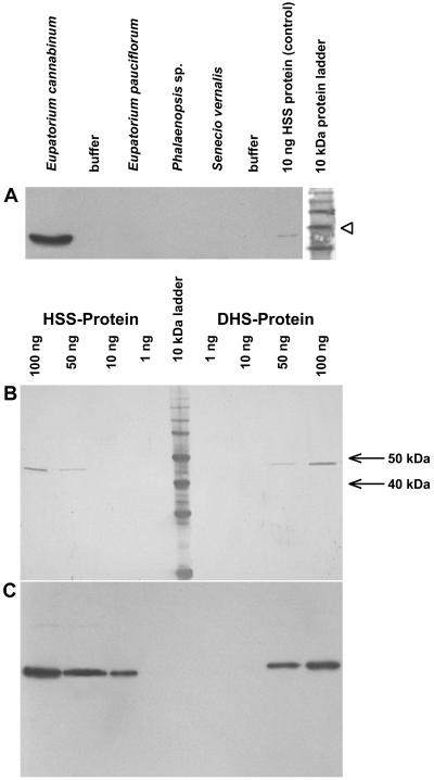
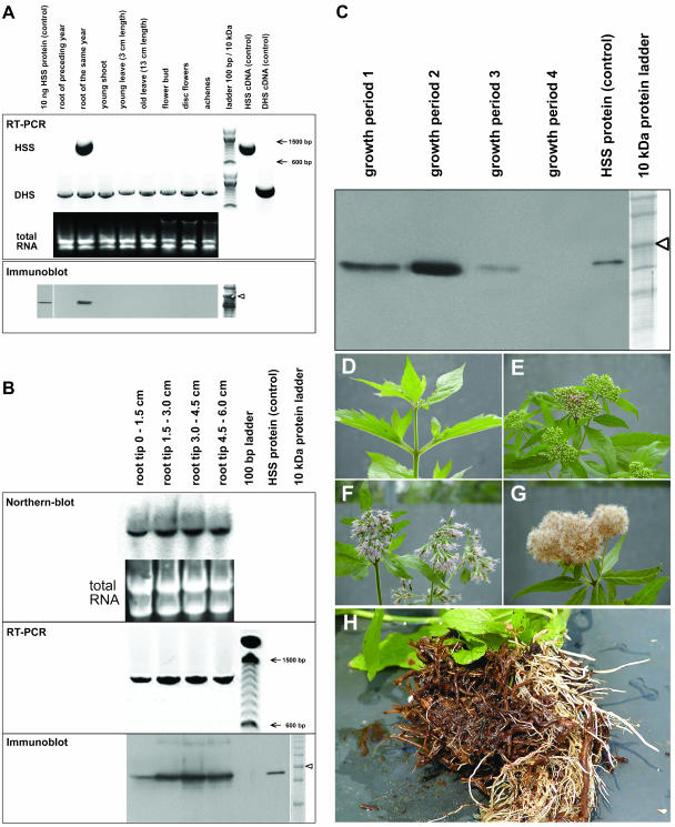
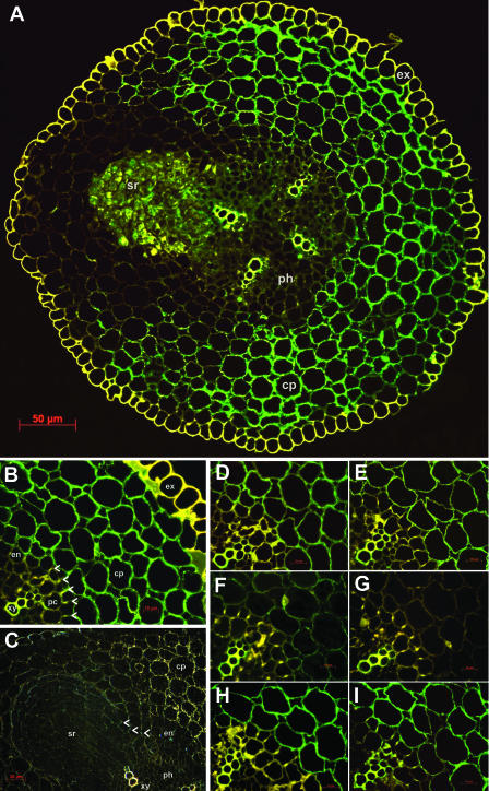
The evolution of pathways within plant secondary metabolism has been studied by using the pyrrolizidine alkaloids (PAs) as a model system. PAs are constitutively produced by plants as a defense against herbivores. The occurrence of PAs is restricted to certain unrelated families within the angiosperms. Homospermidine synthase (HSS), the first specific enzyme in the biosynthesis of the necine base moiety of PAs, was originally recruited from deoxyhypusine synthase, an enzyme involved in the posttranslational activation of the eukaryotic initiation factor 5A. Recently, this gene recruitment has been shown to have occurred several times independently within the angiosperms and even twice within the Asteraceae. Here, we demonstrate that, within these two PA-producing tribes of the Asteraceae, namely Senecioneae and Eupatorieae, HSS is expressed differently despite catalyzing the same step in PA biosynthesis. Within Eupatorium cannabinum, HSS is expressed uniformly in all cells of the root cortex parenchyma, but not within the endodermis and exodermis. Within Senecio vernalis, HSS expression has been previously identified in groups of specialized cells of the endodermis and the adjacent root cortex parenchyma. This expression pattern was confirmed for Senecio jacobaea as well. Furthermore, the expression of HSS in E. cannabinum is dependent on the development of the plant, suggesting a close linkage to plant growth.


Figures

Figure 1.
Figure 1.
Structural types of PAs within the Asteraceae and the reactions catalyzed by HSS and DHS. A, Senecionine N-oxide and lycopsamine N-oxide as typical structures of PAs found within the Senecioneae and Eupatorieae, respectively. The necine base moiety is a bicyclic structure that is common to all PAs. B, HSS and DHS catalyze the transfer of an aminobutyl moiety of spermidine to putrescine (HSS) or to a specific protein-bound Lys residue (DHS).
Figure 2.
Figure 2.
Specificity of the affinity-purified antibody against the HSS of E. cannabinum. A, Immunoblot of proteins extracted from PA-biosynthesizing tissues of various plant species with the antibody specific for HSS of E. cannabinum. Twenty-microgram aliquots of each extract were applied to the gel, and the protein size marker was stained separately with Indian ink. The 50-kD band is indicated by an arrowhead. B, Membranes with identical amounts of pure HSS and DHS after protein staining with Indian ink. C, Membrane identical to B after immunoblot detection with the affinity-purified antibody against the HSS of E. cannabinum.
Figure 3.
Figure 3.
Expression analyses of HSS and DHS in E. cannabinum in various tissues (A), in segments of the root tip (B), and at different seasonal stages (C–G). A, For RT-PCR, cDNAs resulting from reverse transcription of identical amounts of total RNA from various tissues were used as templates for PCR with primers specific for HSS and DHS, respectively. As a control, 50 pg of plasmid DNA carrying the respective cDNAs were coamplified to ensure specificity of the primers used. A 100-bp DNA size marker is shown. For the immunoblot, 20 μg of soluble protein extracted from the same tissues as used for RT-PCR were applied. The 50-kD band of the 10-kD protein size marker is indicated by an arrowhead. B, For northern-blot analysis, total RNA was isolated from 15-mm consecutive segments of the root tip. Ten micrograms of each segment were used as loading control and for hybridization with a probe specific for HSS-coding cDNA. RT-PCR was performed with cDNA from each segment with primers specific for HSS. For immunoblot, proteins were extracted from each segment and detected with the affinity-purified antibody against the HSS of E. cannabinum. The 50-kD band of the 10-kD protein size marker is indicated by an arrowhead. C, Immunoblot of proteins extracted from E. cannabinum roots at different seasonal states of the plant. The 50-kD band of the protein size marker is indicated by an arrowhead. D to G illustrate the growth periods 1 to 4, respectively, as given in C. H, Roots of field-grown E. cannabinum plants with older brown roots and newly grown white roots.
Figure 4.
Figure 4.
Immunolabeling of HSS in root sections of E. cannabinum. A, FITC labeling of HSS within the cortex parenchyma (cp; green). The four xylem rays in the central cylinder and the exodermis (ex) show yellow autofluorescence. A lateral root (sr) is emerging from the pericycle above the xylem ray in the upper left part of the section. B, FITC labeling of HSS in a cutout of A. HSS expression is restricted to cells of the cortex parenchyma (cp); cells of the endodermis (en) and of the exodermis (ex) do not show any label. The casparian strip (arrowheads), the xylem (xy), and the exodermis (ex) show yellow autofluorescence. pc, Pericycle. C, Immunogold detection of the cross-section with the emerging lateral root in detail. Labeled HSS appears as golden grains in the cortex parenchyma (cp); the incrustation of the casparian strip in the radial cell walls of the endodermis (en) shows a faint blue color (indicated by arrowheads). D to I, Cutouts of B that were labeled with HSS-specific antibody according to the standard protocol (D), and after preincubation of the antibody with BSA (E), HSS (F and G), and DHS (H and I) in a molar ratio of antibody to added protein of 10:1 (F and H) and 1:3 (E, G, and I).
Figure 5.
Figure 5.
Electron micrographs of in situ immunogold-labeled HSS in roots of E. cannabinum. A, Cells of the cortex parenchyma (cp) adjoining an intercellular space (ic). cw, Cell wall. B, Two cells of the endodermis (en) separated by a cell wall (cw) carrying the casparian strip (cs), an intercellular (ic), and an adjoining cell of the cortex parenchyma (cp) exclusively being labeled by the HSS-specific antibody.

References

    1. Bayer RJ, Starr JR (1998) Tribal phylogeny of the Asteraceae based on two noncoding chloroplast sequences, the trnL intron and trnL/trnF intergenic spacer. Ann Mo Bot Gard 85: 242–256
    1. Bird DA, Franceschi VR, Facchini PJ (2003) A tale of three cell types: alkaloid biosynthesis is localized to sieve elements in opium poppy. Plant Cell 15: 2626–2635 - PMC - PubMed
    1. Böttcher F, Adolph RD, Hartmann T (1993) Homospermidine synthase, the first pathway-specific enzyme in pyrrolizidine alkaloid biosynthesis. Phytochemistry 32: 679–689 - PMC - PubMed
    1. Böttcher F, Ober D, Hartmann T (1994) Biosynthesis of pyrrolizidine alkaloids: Putrescine and spermidine are essential substrates of enzymatic homospermidine formation. Can J Chem 72: 80–85
    1. Bradford MM (1976) A rapid and sensitive method for the quantitation of microgram quantities of protein utilizing the principle of protein-dye binding. Anal Biochem 72: 248–254 - PubMed

Publication types

MeSH terms

Substances

LinkOut - more resources