. 2004 Dec;136(4):3933-44.
doi: 10.1104/pp.104.051748. Epub 2004 Nov 19.

Plant-specific microtubule-associated protein SPIRAL2 is required for anisotropic growth in Arabidopsis

Affiliations

Plant-specific microtubule-associated protein SPIRAL2 is required for anisotropic growth in Arabidopsis

Tsubasa Shoji et al. Plant Physiol. 2004 Dec.

Erratum in

  • Plant Physiol. 2005 Mar;137(3):1169. Hayashi, Kazuyuki [corrected to Hayashi, Kazunori]

Abstract

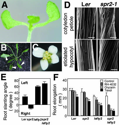
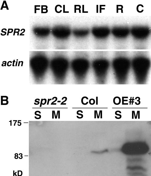
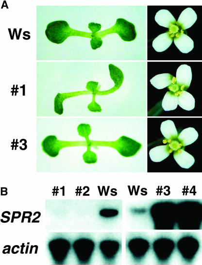
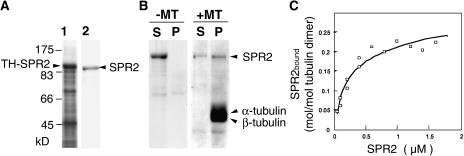
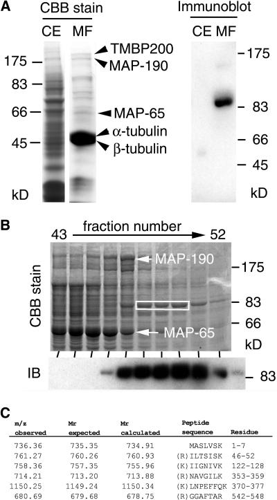
In diffusely growing plant cells, cortical microtubules play an important role in regulating the direction of cell expansion. Arabidopsis (Arabidopsis thaliana) spiral2 (spr2) mutant is defective in directional cell elongation and exhibits right-handed helical growth in longitudinally expanding organs such as root, hypocotyl, stem, petiole, and petal. The growth of spr2 roots is more sensitive to microtubule-interacting drugs than is wild-type root growth. The SPR2 gene encodes a plant-specific 94-kD protein containing HEAT-repeat motifs that are implicated in protein-protein interaction. When expressed constitutively, SPR2-green fluorescent protein fusion protein complemented the spr2 mutant phenotype and was localized to cortical microtubules as well as other mitotic microtubule arrays in transgenic plants. Recombinant SPR2 protein directly bound to taxol-stabilized microtubules in vitro. Furthermore, SPR2-specific antibody and mass spectrometry identified a tobacco (Nicotiana tabacum) SPR2 homolog in highly purified microtubule-associated protein fractions from tobacco BY-2 cell cultures. These results suggest that SPR2 is a novel microtubule-associated protein and is required for proper microtubule function involved in anisotropic growth.


Figures

Figure 1.
Figure 1.
spr2 shows right-handed helical growth and increased sensitivity to microtubule-interacting drugs. A, Cotyledon twisted in a counterclockwise direction in a 10-d-old spr2-1 seedling. B, Rosette leaves twisted in a counterclockwise direction in a 1-month-old spr2-1 plant. Arrowheads indicate the abaxial side of leaves facing up. C, Petals twisted in a counterclockwise direction in a spr2-1 flower. D, Scanning electron micrographs of cotyledon petioles and etiolated hypocotyls of 7-d-old Ler and spr2-1 seedlings. Bars = 100 μm. E, Root-slanting angle of 7-d-old seedlings grown on vertically placed agar plates. Left and right indicate that roots grow toward the left or right side of the plates. F, Inhibition of root growth by microtubule-interacting drugs. After seedlings were grown for 7 d on agar plates containing 0.1 μm oryzalin, 10 nm RH-4032, or 1 μm taxol, lengths of primary roots were measured. More than 30 roots were measured for each treatment and genotype in E and F.
Figure 2.
Figure 2.
Map-based cloning of SPR2. A, Recombination mapping of spr2-1. Mapping with the PCR-based markers localized the SPR2 locus to a region spanned by BACs F10M23, T24A18, and M4I22. B, A schematic diagram of SPR2. SPR2 contains four exons (thick boxes) separated by three introns (lines). The ORF in the exons is shown in black. Mutant lesions are indicated for five spr2 alleles. C, Molecular complementation of spr2. The pBI-SPR2 vector in which SPR2 cDNA was expressed with the 5′ and 3′ regions of the SPR2 gene was introduced into the spr2-2 mutant.
Figure 3.
Figure 3.
SPR2 is a novel, HEAT-repeat-containing protein. A, Phylogenetic tree of SPR2 and its relatives. Full-length amino acid sequences were aligned with the ClustalW method (http://www.ebi.ac.uk/clustalw/#), and the unrooted phylogram was generated using TreeView (http://taxonomy.zoology.gla.ac.uk/rod/treeview.html). The GenBank protein ID number is Q8H513 for the rice SPR2 homolog and CAD45375 for potato HIP2 (Guo et al., 2003). B, Alignment of deduced amino acid sequences of SPR2, its closest Arabidopsis homolog (At1g50890), and potato HIP2. Residues identical in a least two sequences are shaded in gray, and dashes indicate gaps introduced to maximize the alignment. Nine HEAT-repeat motifs are underlined. Asterisks above the N-terminal sequences (amino acids 1–37 in SPR2) indicate Ser and Thr residues. Peptide fragments of HIP2 that were identified by GC-MS/MS analysis in Figure 8C are boxed. C, Alignment of nine predicted HEAT-repeat motifs in SPR2. HEAT-repeat motifs consist of two α-helices containing moderately conserved amino acid residues at the indicated positions (Andrade et al., 2001). Residues conforming to the consensus are shaded in black, and similar residues are shaded in gray. Gaps were introduced in the hinge region between the two α-helices to maximize the alignment.
Figure 4.
Figure 4.
SPR2 expression and protein accumulation. A, RT-PCR analysis of SPR2 expression in various tissues of wild-type Col plants. FB, flower bud; CL, cauline leaf; RL, rosette leaf; IF, inflorescence stem; R, root; C, cotyledon. Actin gene expression was used as a control. B, Immunoblot analysis with SPR2-specific antibody. Soluble and microsomal proteins (10 μg each) were analyzed in 7-d-old seedlings of Col, spr2-2, and a SPR2-overexpressing line (OE 3).
Figure 5.
Figure 5.
SPR2 overexpression and cosuppression. SPR2 cDNA was expressed under the control of the CaMV 35S promoter in Wassilewskija plants. Transgenic lines were numbered from 1 to 4. A, Seedling and flower phenotypes. B, RT-PCR analysis of SPR2 expression in 7-d-old seedlings. Lines 1 and 2 are cosuppression lines, whereas lines 3 and 4 are overexpression lines. Actin gene expression was used as a control.
Figure 6.
Figure 6.
Intracellular localization of SPR2-GFP protein in vivo. SPR2-GFP fusion protein was stably expressed under the control of the CaMV 35S promoter in spr2-2 plants and the spr2 mutant phenotype was rescued in the transgenic plants. Seven-day-old Arabidopsis seedlings were analyzed with confocal microscopy. A, Leaf epidermis. B, Leaf epidermis with a focal plane adjusted to the middle of the cells' thickness. C, Magnified picture of a portion of the leaf epidermal cells shown in A. D, Leaf trichome. E, Epidermis of etiolated hypocotyl. F, Root epidermis at the differentiation zone. G, Effect of propyzamide on the SPR2-GFP localization. Hypocotyls of transgenic seedlings were treated with propyzamide at 50 μm for 1 h. H, Root meristematic region. Mitotic spindles (sp) and phragmoplasts (ph) were labeled with SPR2-GFP. Bars = 25 μm in A and B, 5 μm in C, and 10 μm in D to H.
Figure 7.
Figure 7.
Recombinant SPR2 binds to taxol-stabilized microtubules in vitro. A, Bacterial expression and purification of recombinant SPR2 protein. Full-length SPR2 was fused with thioredoxin and poly-His tags (TH-SPR2), overexpressed in E. coli., and purified to near homogeneity after enzymatic removal of the tags. Crude soluble extract of E. coli and purified SPR2 (without tags) were separated by SDS-PAGE and stained with Coomassie Brilliant Blue. B, Cosedimentation of recombinant SPR2 with taxol-stabilized microtubules. Taxol-stabilized microtubules were prepared from purified tobacco BY-2 tubulins. After recombinant SPR2 was incubated with or without taxol-stabilized microtubules, proteins were centrifuged and analyzed by SDS-PAGE and Coomassie Brilliant Blue staining. The positions of SPR2, α-tubulin, and β-tubulin are indicated by arrowheads. S, Supernatant fraction; P, pellet fraction; and ±MT, presence or absence of microtubules in the assay mixture. C, Quantitative analysis of the binding between SPR2 and microtubules. Various concentrations of purified SPR2 were mixed with taxol-stabilized microtubules (6 μg) and then subjected to cosedimentation analysis, as in B. Assuming there is one SPR2-binding site on the tubulin dimer, the equation q = (qmax × c)/(Kd + c) was fitted. In this equation, q is the amount of protein bound to the tubulin dimer and c is the concentration of free SPR2 in solution. Kd and qmax, which represent the dissociation constant and the amount of bound SPR2 at the saturated level, were calculated from the fitting.
Figure 8.
Figure 8.
Immunological and mass spectrometric identification of tobacco SPR2 homolog in MAP fractions purified from tobacco BY-2 cells. A, Crude protein extract from tobacco miniprotoplasts (CE) and proteins in a purified MAP fraction (MF) were separated by SDS-PAGE and stained with Coomassie Brilliant Blue (left) or subjected to immunoblotting with an anti-SPR2 antibody (right). Arrowheads on the left indicate the positions of TMBP200, MAP-190, MAP-65, α-tubulin, and β-tubulin. B, After proteins in the tobacco MAP fraction were fractionated with anion-exchange chromatography, eluted fractions (43–52) were separated by SDS-PAGE and stained with Coomassie Brilliant Blue (upper image) or analyzed by immunoblotting with an anti-SPR2 antibody (IB; lower image). The positions of MAP-190 and MAP-65 are indicated by arrows. Tobacco SPR2-related protein was abundant in fractions 47 to 51. Coomassie Brilliant Blue-stained bands containing the SPR2-related protein in fractions 48 to 50 (white box) were excised and subjected to LC-MS/MS analysis. C, Peptide sequences identified by LC-MS/MS analysis. Mass values of precursor ions (m/z observed), expected peptide mass values (molecular mass expected), and mass values calculated from the peptide sequences in the database (molecular mass calculated) are shown. Amino acid residues of the identified peptides are numbered based on the HIP2 sequence and are also shown in boxes in Figure 3B. All peptides had an Arg or a Lys at the last residue position and at the position immediately prior to the initial peptide residues in the intact HIP2 sequence (except the N-terminal peptide), as expected for peptides digested with trypsin.

Similar articles

Cited by

References

    1. Abe T, Thitamadee S, Hashimoto T (2004) Microtubule defects and cell morphogenesis in the lefty1lefty2 tubulin mutant of Arabidopsis thaliana. Plant Cell Physiol 45: 211–220 - PubMed
    1. An G (1987) Binary Ti vectors for plant transformation and promoter analysis. Methods Enzymol 153: 292–305
    1. Andrade MA, Bork P (1995) HEAT repeats in the Huntington's disease protein. Nat Genet 11: 115–116 - PubMed
    1. Andrade MA, Petosa C, O'Donoghue SI, Müller CW, Bork P (2001) Comparison of ARM and HEAT protein repeats. J Mol Biol 309: 1–18 - PubMed
    1. Andrade MA, Ponting CP, Gibson TJ, Bork P (2000) Homology-based method for identification of protein repeats using statistical significance estimates. J Mol Biol 298: 521–537 - PubMed

Publication types

MeSH terms

Substances