

Progression through meiosis I and meiosis II in Arabidopsis anthers is regulated by an A-type cyclin predominately expressed in prophase I
- PMID: 15557098
- PMCID: PMC535843
- DOI: 10.1104/pp.104.051201
Progression through meiosis I and meiosis II in Arabidopsis anthers is regulated by an A-type cyclin predominately expressed in prophase I
Abstract
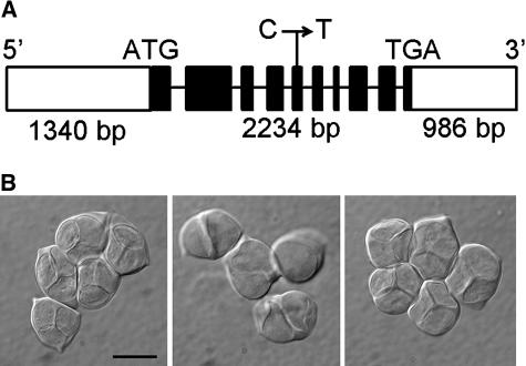
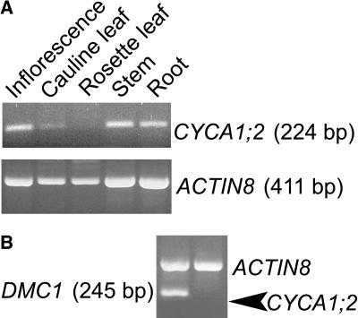
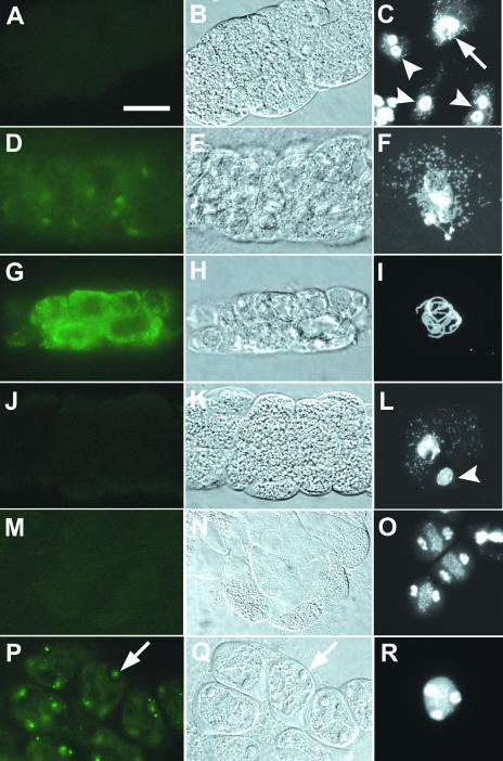
Meiosis is often described as a special case of cell division since it differs from mitosis in having two nuclear divisions without an intervening S-phase. It will be of great interest to uncover what molecular mechanisms underlie these special features of meiosis. We previously reported that the tardy asynchronous meiosis (tam) mutant of Arabidopsis (Arabidopsis thaliana) is slower in cell cycle progression in male meiosis. Here we report that TAM encodes the A-type cyclin, CYCA1;2. The point mutation in tam replaced a conserved threonine with an isoleucine in the linker region between the alpha4 and alpha5 helices of the first cyclin fold. By studying the dynamics of a CYCA1;2-green fluorescent protein fusion protein under the control of the CYCA1;2 promoter, we found that the fusion protein was most abundant at pachytene, but was undetectable from late prophase I until telophase II. Nonetheless, cell cycle progression in tam was delayed in both pachytene and meiosis II. We conclude either that the CYCA1;2 produced in prophase I indirectly regulates meiosis II progression, or that a very low level of CYCA1;2 directly regulates meiosis II progression. Either of these scenarios is a deviation from the typical mode of action of mitotic cyclins in mitosis and meiosis I, in which each nuclear division is coupled with a peak of expression of mitotic cyclins.
Figures


Similar articles
-
The cyclin-A CYCA1;2/TAM is required for the meiosis I to meiosis II transition and cooperates with OSD1 for the prophase to first meiotic division transition.PLoS Genet. 2010 Jun 17;6(6):e1000989. doi: 10.1371/journal.pgen.1000989. PLoS Genet. 2010. PMID: 20585549 Free PMC article.
-
Loss-of-function mutants and overexpression lines of the Arabidopsis cyclin CYCA1;2/Tardy Asynchronous Meiosis exhibit different defects in prophase-i meiocytes but produce the same meiotic products.PLoS One. 2014 Nov 17;9(11):e113348. doi: 10.1371/journal.pone.0113348. eCollection 2014. PLoS One. 2014. PMID: 25402453 Free PMC article.
-
OSD1 promotes meiotic progression via APC/C inhibition and forms a regulatory network with TDM and CYCA1;2/TAM.PLoS Genet. 2012;8(7):e1002865. doi: 10.1371/journal.pgen.1002865. Epub 2012 Jul 26. PLoS Genet. 2012. PMID: 22844260 Free PMC article.
-
Regulating mitosis and meiosis in the male germ line: critical functions for cyclins.Philos Trans R Soc Lond B Biol Sci. 2010 May 27;365(1546):1653-62. doi: 10.1098/rstb.2009.0254. Philos Trans R Soc Lond B Biol Sci. 2010. PMID: 20403876 Free PMC article. Review.
-
Genetic Regulation of Mitosis-Meiosis Fate Decision in Plants: Is Callose an Oversighted Polysaccharide in These Processes?Plants (Basel). 2023 May 9;12(10):1936. doi: 10.3390/plants12101936. Plants (Basel). 2023. PMID: 37653853 Free PMC article. Review.
Cited by
-
The cyclin-A CYCA1;2/TAM is required for the meiosis I to meiosis II transition and cooperates with OSD1 for the prophase to first meiotic division transition.PLoS Genet. 2010 Jun 17;6(6):e1000989. doi: 10.1371/journal.pgen.1000989. PLoS Genet. 2010. PMID: 20585549 Free PMC article.
-
Characterization of a set of novel meiotically-active promoters in Arabidopsis.BMC Plant Biol. 2012 Jul 9;12:104. doi: 10.1186/1471-2229-12-104. BMC Plant Biol. 2012. PMID: 22776406 Free PMC article.
-
SUMO E3 ligase AtMMS21 is required for normal meiosis and gametophyte development in Arabidopsis.BMC Plant Biol. 2014 Jun 3;14:153. doi: 10.1186/1471-2229-14-153. BMC Plant Biol. 2014. PMID: 24893774 Free PMC article.
-
The Chromatin Protein DUET/MMD1 Controls Expression of the Meiotic Gene TDM1 during Male Meiosis in Arabidopsis.PLoS Genet. 2015 Sep 8;11(9):e1005396. doi: 10.1371/journal.pgen.1005396. eCollection 2015 Sep. PLoS Genet. 2015. PMID: 26348709 Free PMC article.
-
Meiotic progression in Arabidopsis is governed by complex regulatory interactions between SMG7, TDM1, and the meiosis I-specific cyclin TAM.Plant Cell. 2010 Nov;22(11):3791-803. doi: 10.1105/tpc.110.078378. Epub 2010 Nov 30. Plant Cell. 2010. PMID: 21119056 Free PMC article.
References
-
- Armstrong SJ, Jones GH (2003) Meiotic cytology and chromosome behavior in wild-type Arabidopsis thaliana. J Exp Bot 54: 1–10 - PubMed
-
- Buonomo SBC, Rabitsch KP, Fuchs J, Gruber S, Sullivan M, Uhlmann F, Petronczki M, Toth A, Nasmyth K (2003) Division of the nucleolus and its release of CDC14 during anaphase of meiosis I depends on separase, SPO12, and SLK19. Dev Cell 4: 727–739 - PubMed
-
- Caryl AP, Armstrong SJ, Jones GH, Franklin FC (2000) A homologue of the yeast HOP1 gene is inactivated in the Arabidopsis meiotic mutant asy1. Chromosoma 109: 62–71 - PubMed
Publication types
MeSH terms
Substances
LinkOut - more resources
Full Text Sources
Other Literature Sources
Molecular Biology Databases
