Molecular modeling of family GH16 glycoside hydrolases: potential roles for xyloglucan transglucosylases/hydrolases in cell wall modification in the poaceae
- PMID: 15557263
- PMCID: PMC2287310
- DOI: 10.1110/ps.04828404
Molecular modeling of family GH16 glycoside hydrolases: potential roles for xyloglucan transglucosylases/hydrolases in cell wall modification in the poaceae
Abstract
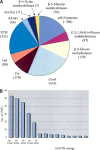
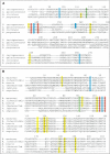
Family GH16 glycoside hydrolases can be assigned to five subgroups according to their substrate specificities, including xyloglucan transglucosylases/hydrolases (XTHs), (1,3)-beta-galactanases, (1,4)-beta-galactanases/kappa-carrageenases, "nonspecific" (1,3/1,3;1,4)-beta-D-glucan endohydrolases, and (1,3;1,4)-beta-D-glucan endohydrolases. A structured family GH16 glycoside hydrolase database has been constructed (http://www.ghdb.uni-stuttgart.de) and provides multiple sequence alignments with functionally annotated amino acid residues and phylogenetic trees. The database has been used for homology modeling of seven glycoside hydrolases from the GH16 family with various substrate specificities, based on structural coordinates for (1,3;1,4)-beta-D-glucan endohydrolases and a kappa-carrageenase. In combination with multiple sequence alignments, the models predict the three-dimensional (3D) dispositions of amino acid residues in the substrate-binding and catalytic sites of XTHs and (1,3/1,3;1,4)-beta-d-glucan endohydrolases; there is no structural information available in the databases for the latter group of enzymes. Models of the XTHs, compared with the recently determined structure of a Populus tremulos x tremuloides XTH, reveal similarities with the active sites of family GH11 (1,4)-beta-D-xylan endohydrolases. From a biological viewpoint, the classification, molecular modeling and a new 3D structure of the P. tremulos x tremuloides XTH establish structural and evolutionary connections between XTHs, (1,3;1,4)-beta-D-glucan endohydrolases and xylan endohydrolases. These findings raise the possibility that XTHs from higher plants could be active not only on cell wall xyloglucans, but also on (1,3;1,4)-beta-D-glucans and arabinoxylans, which are major components of walls in grasses. A role for XTHs in (1,3;1,4)-beta-D-glucan and arabinoxylan modification would be consistent with the apparent overrepresentation of XTH sequences in cereal expressed sequence tags databases.
Figures


Similar articles
-
Analysis of nasturtium TmNXG1 complexes by crystallography and molecular dynamics provides detailed insight into substrate recognition by family GH16 xyloglucan endo-transglycosylases and endo-hydrolases.Proteins. 2009 Jun;75(4):820-36. doi: 10.1002/prot.22291. Proteins. 2009. PMID: 19004021
-
Crystallographic insight into the evolutionary origins of xyloglucan endotransglycosylases and endohydrolases.Plant J. 2017 Feb;89(4):651-670. doi: 10.1111/tpj.13421. Epub 2017 Feb 11. Plant J. 2017. PMID: 27859885 Free PMC article.
-
Structure-function analysis of a broad specificity Populus trichocarpa endo-β-glucanase reveals an evolutionary link between bacterial licheninases and plant XTH gene products.J Biol Chem. 2013 May 31;288(22):15786-99. doi: 10.1074/jbc.M113.462887. Epub 2013 Apr 9. J Biol Chem. 2013. PMID: 23572521 Free PMC article.
-
Structure-function relationships of beta-D-glucan endo- and exohydrolases from higher plants.Plant Mol Biol. 2001 Sep;47(1-2):73-91. Plant Mol Biol. 2001. PMID: 11554481 Review.
-
Polysaccharide hydrolases in germinated barley and their role in the depolymerization of plant and fungal cell walls.Int J Biol Macromol. 1997 Aug;21(1-2):67-72. doi: 10.1016/s0141-8130(97)00043-3. Int J Biol Macromol. 1997. PMID: 9283018 Review.
Cited by
-
Discovery of small molecule inhibitors of xyloglucan endotransglucosylase (XET) activity by high-throughput screening.Phytochemistry. 2015 Sep;117:220-236. doi: 10.1016/j.phytochem.2015.06.016. Epub 2015 Jun 19. Phytochemistry. 2015. PMID: 26093490 Free PMC article.
-
Polysaccharide microarrays for high-throughput screening of transglycosylase activities in plant extracts.Glycoconj J. 2010 Jan;27(1):79-87. doi: 10.1007/s10719-009-9271-8. Epub 2009 Dec 2. Glycoconj J. 2010. PMID: 19953317
-
Considerations in the search for mixed-linkage (1→3),(1→4)-β-D-glucan-active endotransglycosylases.Plant Signal Behav. 2013 Apr;8(4):e23835. doi: 10.4161/psb.23835. Epub 2013 Feb 20. Plant Signal Behav. 2013. PMID: 23425852 Free PMC article.
-
DWARF--a data warehouse system for analyzing protein families.BMC Bioinformatics. 2006 Nov 9;7:495. doi: 10.1186/1471-2105-7-495. BMC Bioinformatics. 2006. PMID: 17094801 Free PMC article.
-
Plant cell wall integrity maintenance in model plants and crop species-relevant cell wall components and underlying guiding principles.Cell Mol Life Sci. 2020 Jun;77(11):2049-2077. doi: 10.1007/s00018-019-03388-8. Epub 2019 Nov 28. Cell Mol Life Sci. 2020. PMID: 31781810 Free PMC article. Review.
References
-
- Anderson, M.A. and Stone, B.A. 1975. A new substrate for investigating the specificity of β-d-glucan hydrolases. FEBS Lett. 52 202–207. - PubMed
-
- Barbeyron, T., Gerard, A., Potin, P., Henrissat, B., and Kloareg, B. 1998. The κ-carrageenase of the marine bacterium Cytophaga drobachiensis: Structural and phylogenetic relationships within family-16 glycoside hydrolases. Mol. Biol. Evol. 15 528–537. - PubMed
Publication types
MeSH terms
Substances
LinkOut - more resources
Full Text Sources
Other Literature Sources

