. 2004 Dec 6;200(11):1479-89.
doi: 10.1084/jem.20041690. Epub 2004 Nov 22.

Cytolytic CD8+ T cells recognizing CFP10 are recruited to the lung after Mycobacterium tuberculosis infection

Affiliations

Cytolytic CD8+ T cells recognizing CFP10 are recruited to the lung after Mycobacterium tuberculosis infection

Arati B Kamath et al. J Exp Med. .

Abstract

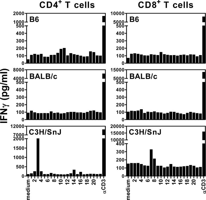
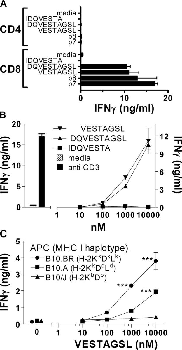
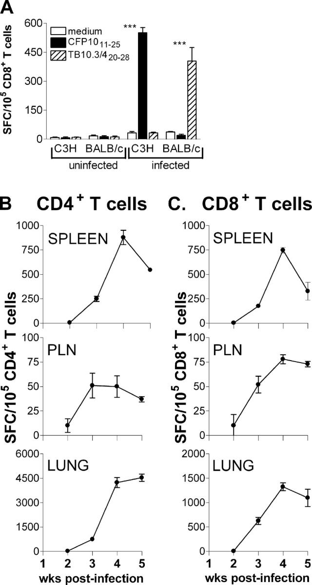
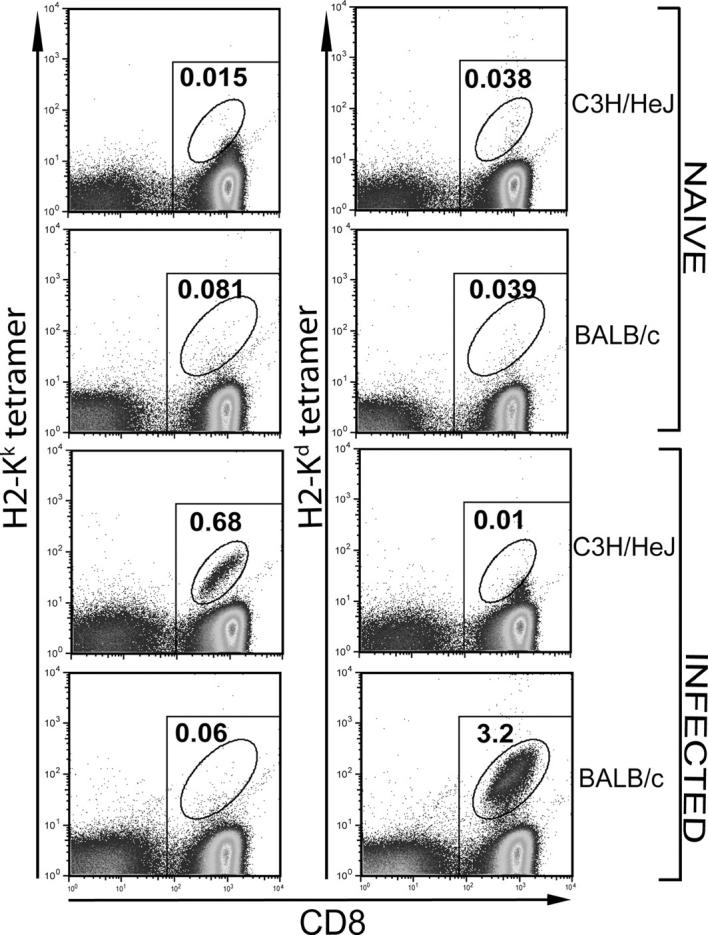
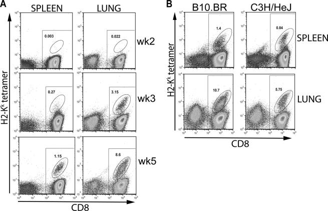
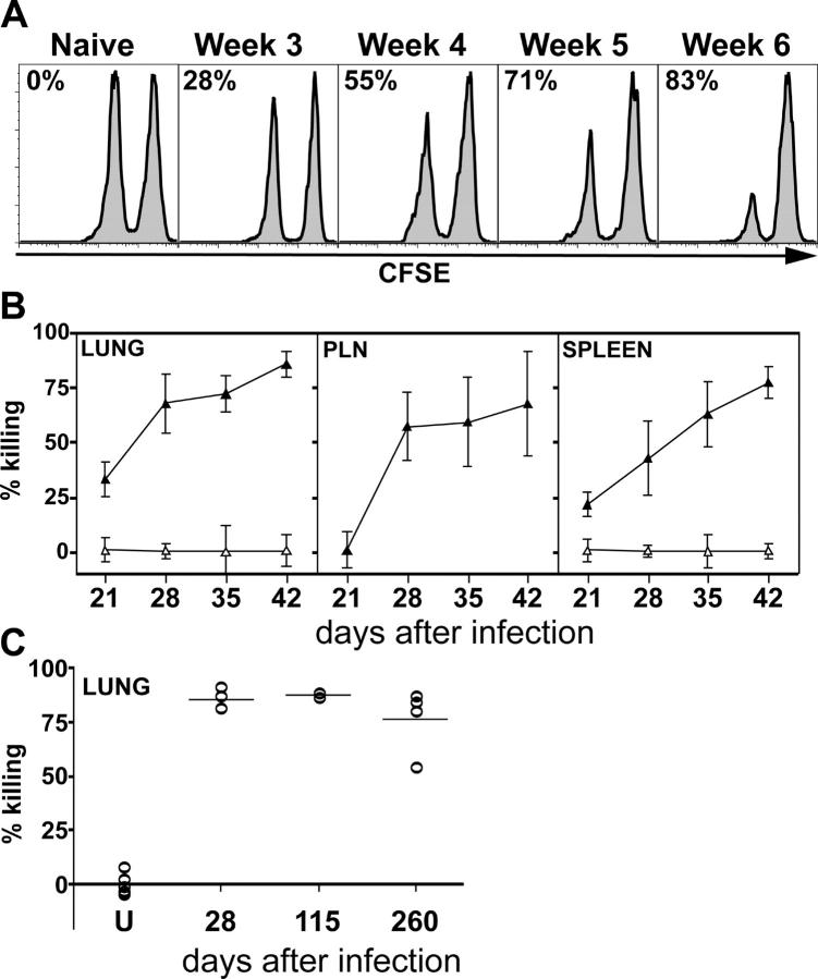
Optimum immunity against Mycobacterium tuberculosis requires both CD4(+) and CD8(+) T cells. In contrast with CD4(+) T cells, few antigens are known that elicit CD8(+) T cells during infection. CD8(+) T cells specific for culture filtrate protein-10 (CFP10) are found in purified protein derivative positive donors, suggesting that CFP10 primes CD8(+) T cells in vivo. Using T cells from M. tuberculosis-infected mice, we identified CFP10 epitopes recognized by CD8(+) T cells and CD4(+) T cells. CFP10-specific T cells were detected as early as week 3 after infection and at their peak accounted for up to 30% of CD8(+) T cells in the lung. IFNgamma-producing CD8(+) and CD4(+) T cells recognizing CFP10 epitopes were preferentially recruited to the lungs of M. tuberculosis-infected mice. In vivo cytolytic activity of CD8(+) T cells specific for CFP10 and TB10.3/10.4 proteins was detected in the spleen, pulmonary lymph nodes, and lungs of infected mice. The cytolytic activity persisted long term and could be detected 260 d after infection. This paper highlights the cytolytic function of antigen-specific CD8(+) T cells elicited by M. tuberculosis infection and demonstrates that large numbers of CFP10-specific cytolytic CD8(+) T cells are recruited to the lung after M. tuberculosis infection.


Figures

Figure 1.
Figure 1.
Identification of a new MHC class I–restricted M. tuberculosis epitope recognized by CD8+ T cells. Purified CD4+ and CD8+ T cells from splenocytes of week 4–infected C57BL/6, BALB/c, and C3H/HeSnJ mice were cultured with CFP10 peptides at a concentration of 10 μM and syngeneic naive irradiated splenocytes as APCs. The amount of IFNγ released into the supernatants was measured by ELISA after 48 h. The data represent one out of three independent experiments with similar results
Figure 2.
Figure 2.
Identification of the minimal epitope and restricting element recognized by CD8+ T cells. (A) Definition of the minimal epitope recognized by CFP10-specific CD8+ T cells. Purified CD4+ and CD8+ splenic T cells obtained from C3H/HeSnJ mice 4 wk after infection were cultured with p7 or p8 from the CFP10 peptide library, or the overlapping peptides VESTAGSL, DQVESTAGSL, and IDQVESTA (all at 10 μM). IFNγ in the culture supernatants was measured after 48 h by ELISA. The CD8+ T cell responses to VESTAGSL, DQVESTAGSL, p7, and p8 are all statistically significant (P < 0.05) compared with media alone when analyzed by one-way ANOVA. (B) Dose–response of CFP10-specific CD8+ T cells to specific peptides. Splenic CD8+ T cell responses to overlapping peptides VESTAGSL, DQVESTAGSL, and IDQVESTA. The responses to VESTAGSL and DQVESTAGSL were similar to each other and both are significantly different from the response to IDQVESTA (P < 0.01, by two-way ANOVA). (C) Recognition of CFP10 by CD8+ T cells was restricted by H-2Kk. IFNγ production was used to indicate splenic CD8+ T cell recognition of the VESTAGSL peptide when presented by irradiated splenocytes obtained from B10.BR, B10.A, or B10.J mice. ***, P < 0.001 by two-way ANOVA with Bonferroni's posttest. Results shown are representative of at least three independent experiments. Error bars represent SD.
Figure 3.
Figure 3.
Enumeration of the peptide-specific CD4+ and CD8+ T cells after M. tuberculosis infection using the ELISPOT assay. (A) Epitope specificity and H-2 restriction of the ELISPOT assay. Purified splenic CD8+ T cells from uninfected or infected C3H/HeJ or BALB/c mice were cultured with syngeneic APCs in the presence of the H-2Kk–restricted CFP1032-39 or H-2Kd–restricted TB10.3/420-28 peptides and an IFNγ ELISPOT assay was performed. ***, P < 0.001 by two-way ANOVA with Bonferroni's posttest. (B and C) The kinetics of the CFP10-specific T cell response. Purified CD4+ T cells (B) or CD8+ T cells (C) from infected C3H/HeJ mice were cultured with the I-Ak–restricted CFP1011-25 peptide or the H-2Kk–restricted CFP1032-39 peptide in the presence of syngeneic irradiated splenocytes as APCs. The T cells were purified from pooled organs of six infected mice at each time point. Controls without peptide produced a minimal background level of IFNγ+ spots and this was subtracted from the number of spots in the presence of the peptide. The data are representative of two independent experiments and shown as the number of spot-forming cells/100,000 CD4+ or CD8+ T cells. Error bars represent SD.
Figure 4.
Figure 4.
H-2Kk and H-2Kd peptide tetramer staining of CD8+ T cells in M. tuberculosis–infected C3H/HeJ or BALB/c mice. Purified CD8+ T cells from splenocytes of naive or 4 wk–infected C3H/HeJ or BALB/c mice were stained with the CFP1032-39/H-2Kk or TB10.3/420-28/H-2Kd tetramers. A significant population of H-2Kk and the H-2Kd tetramer+ CD8+ T cells was found only in cells obtained from the infected C3H/HeJ and BALB/c mice, respectively. All of the CD8+ T cells are within the rectangular gate and all of the tetramer+ cells are within the oval gate. The number represents the percentage of CD8+ T cells that are tetramer+.
Figure 5.
Figure 5.
Detection of tetramer+ CD8+ T cells in the spleens and lungs of C3H/HeJ and B10.BR mice after M. tuberculosis infection. (A) Purified CD8+ T cells from splenocytes of infected C3H/HeJ mice were stained at various times after infection. Tetramer+ CD8+ T cells could be detected in the spleens and lungs of infected C3H/HeJ mice as early as 3 wk after infection. The percent of tetramer+ CD8+ T cells is shown in all panels. (B) CFP10-specific CD8+ T cells in C3H/HeJ and B10.BR mice after infection. The percentage of tetramer+ CD8+ T cells found in the spleens and lungs of resistant B10.BR mice and susceptible C3H/HeJ mice were compared 4 wk after infection with M. tuberculosis. All of the CD8+ T cells are within the rectangular gate and all of the tetramer+ cells are within the oval gate. The number represents the percentage of CD8+ T cells that are tetramer+.
Figure 6.
Figure 6.
Generation of peptide-specific cytotoxic CD8+ T cells after M. tuberculosis infection. (A) In vivo cytolytic activity of CFP1032-39-specific CD8+ T cells. A 1:1 mixture of CFSEhigh and CFP1032-39-pulsed CFSElow splenocytes were injected i.v. into C3H/HeJ that were uninfected or infected 3–6 wk previously with M. tuberculosis. The spleens of the recipient mice were removed after 18 h and analyzed by flow cytometry. A histogram gated on CFSE+ events is shown for a representative mouse from each group. The number in each plot represents the percent killing of the CFP1032-39-pulsed CFSElow splenocytes, which was calculated as described in Materials and Methods. (B) The relative killing of the peptide-pulsed targets in the lungs, PLNs, and spleens of infected mice compared with uninfected mice was calculated as described in Materials and Methods. Each point represents the mean ± SD of five mice per group. The experiment was repeated twice with similar results. (C) Uninfected (U) or infected BALB/c mice were injected as described before with TB10.3/420-28-pulsed and unpulsed splenocytes. The percent killing in the lungs of individual mice from 28 to 260 after infection is shown. Each symbol represents an individual mouse.

References

    1. Flynn, J.L., and J. Chan. 2001. Immunology of tuberculosis. Annu. Rev. Immunol. 19:93–129. - PubMed
    1. Feng, C.G., A.G. Bean, H. Hooi, H. Briscoe, and W.J. Britton. 1999. Increase in gamma interferon-secreting CD8(+), as well as CD4(+), T cells in lungs following aerosol infection with Mycobacterium tuberculosis. Infect. Immun. 67:3242–3247. - PMC - PubMed
    1. Smith, S.M., and H.M. Dockrell. 2000. Role of CD8 T cells in mycobacterial infections. Immunol. Cell Biol. 78:325–333. - PubMed
    1. Ackerman, A.L., and P. Cresswell. 2004. Cellular mechanisms governing cross-presentation of exogenous antigens. Nat. Immunol. 5:678–684. - PubMed
    1. Flynn, J.L., M.M. Goldstein, K.J. Triebold, B. Koller, and B.R. Bloom. 1992. Major histocompatibility complex class I-restricted T cells are required for resistance to Mycobacterium tuberculosis infection. Proc. Natl. Acad. Sci. USA. 89:12013–12017. - PMC - PubMed

Publication types

Substances