. 2004 Dec;72(12):6780-9.
doi: 10.1128/IAI.72.12.6780-6789.2004.

Yersinia enterocolitica adhesin A induces production of interleukin-8 in epithelial cells

Affiliations

Yersinia enterocolitica adhesin A induces production of interleukin-8 in epithelial cells

Yvonne Schmid et al. Infect Immun. 2004 Dec.

Abstract

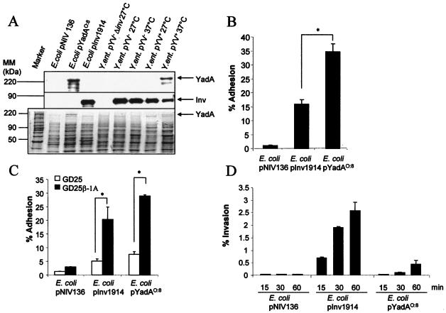
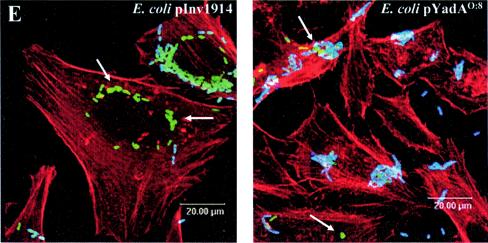
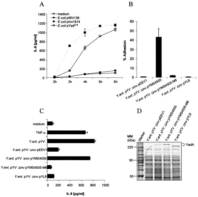
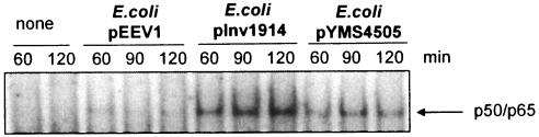
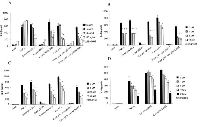
The major invasive factor of Yersinia enterocolitica, the invasin (Inv) protein, induces proinflammatory host cell responses, including interleukin-8 (IL-8) secretion from human epithelial cells, by engagement of beta1 integrins. The Inv-triggered beta1 integrin signaling involves the small GTPase Rac; the activation of MAP kinases, such as p38, MEK1, and JNK; and the activation of the transcription factor NF-kappaB. In the present study, we demonstrate that Y. enterocolitica YadA, which is a major adhesin of Y. enterocolitica with pleiotropic virulence effects, induces IL-8 secretion in epithelial cells. The abilities of YadA and Inv to promote adhesion to and invasion of HeLa cells and to induce IL-8 production by the cells were investigated by expression of YadA and Inv in Escherichia coli. While YadA mediates efficacious adhesion to HeLa cells, it mediates marginal invasion compared with Inv. Both YadA and Inv trigger comparable levels of IL-8 production. Conformational changes of the YadA head domain by mutation of NSVAIG-S motifs, which abolish collagen binding, also abolish adhesion of Yersinia to HeLa cells and YadA-mediated IL-8 secretion. Furthermore, experiments in which blocking antibodies against beta1 integrins were used demonstrate that beta1 integrins are crucial for YadA-mediated IL-8 secretion. Inhibitor studies demonstrate the involvement of small GTPases and MAP kinases, such as p38, MEK1, and JNK, indicating that beta1 integrin-dependent signaling mediated by Inv or YadA involves similar signaling pathways. These data present YadA, in addition to Inv, YopB, and Yersinia lipopolysaccharide, as a further inducer of proinflammatory molecules by which Y. enterocolitica might promote inflammatory tissue reactions.


Figures

FIG. 1.
FIG. 1.
YadA- and Inv-mediated adhesion and invasion. (A) Expression of YadA and Inv of E. coli pYadAO:8, E. coli pInv1914, Y. enterocolitica pYV+, and Y. enterocolitica pYV. Yersinia and E. coli strains were grown overnight in LB at 27 and 37°C and subcultured for a further 3 h at the indicated temperatures. Subsequently, Western blot analysis using polyclonal antibodies against Inv (middle) and YadA (top) was performed. In parallel, SDS-PAGE (bottom) was performed as a loading control. Inv and YadA expression is indicated by arrows. (B) HeLa cells were infected with Inv- or YadA-expressing bacteria (MOI, 100). Adhesion of bacteria to HeLa cells was determined 15 min postinfection, as described in Materials and Methods. The asterisk indicates a significant difference between adhesion of E. coli pYadAO:8 and that of E. coli pInv1914 to HeLa cells (P < 0.05). The error bars indicate standard deviations. (C) GD25 cells were infected with Inv- or YadA-expressing bacteria (MOI, 100). Adhesion of bacteria to GD25-β1A and GD25 cells was determined 15 min postinfection. The asterisks indicate a significant difference between the adhesion of E. coli pINV1914 and that of E. coli YadAO:8 to GD25 cells versus GD25-β1A cells (P < 0.05). (D) Uptake of bacteria into HeLa cells was analyzed by determining the number of intracellularly viable bacteria using gentamicin killing at the indicated time points. (E) Internalization of E. coli pInv1914 or E. coli pYadAO:8 into HeLa cells was visualized by immunofluorescence using polyclonal antibodies against Inv or YadA, respectively, as indicated in Material and Methods. Overlays of multicolor staining are shown. The cytoskeleton was stained with phalloidin-tetramethyl rhodamine isothiocyanate (red). Extracellularly localized bacteria, blue and green staining; internalized bacteria, exclusively green staining. The arrows indicate examples of intracellularly located bacteria.
FIG. 1.
FIG. 1.
YadA- and Inv-mediated adhesion and invasion. (A) Expression of YadA and Inv of E. coli pYadAO:8, E. coli pInv1914, Y. enterocolitica pYV+, and Y. enterocolitica pYV. Yersinia and E. coli strains were grown overnight in LB at 27 and 37°C and subcultured for a further 3 h at the indicated temperatures. Subsequently, Western blot analysis using polyclonal antibodies against Inv (middle) and YadA (top) was performed. In parallel, SDS-PAGE (bottom) was performed as a loading control. Inv and YadA expression is indicated by arrows. (B) HeLa cells were infected with Inv- or YadA-expressing bacteria (MOI, 100). Adhesion of bacteria to HeLa cells was determined 15 min postinfection, as described in Materials and Methods. The asterisk indicates a significant difference between adhesion of E. coli pYadAO:8 and that of E. coli pInv1914 to HeLa cells (P < 0.05). The error bars indicate standard deviations. (C) GD25 cells were infected with Inv- or YadA-expressing bacteria (MOI, 100). Adhesion of bacteria to GD25-β1A and GD25 cells was determined 15 min postinfection. The asterisks indicate a significant difference between the adhesion of E. coli pINV1914 and that of E. coli YadAO:8 to GD25 cells versus GD25-β1A cells (P < 0.05). (D) Uptake of bacteria into HeLa cells was analyzed by determining the number of intracellularly viable bacteria using gentamicin killing at the indicated time points. (E) Internalization of E. coli pInv1914 or E. coli pYadAO:8 into HeLa cells was visualized by immunofluorescence using polyclonal antibodies against Inv or YadA, respectively, as indicated in Material and Methods. Overlays of multicolor staining are shown. The cytoskeleton was stained with phalloidin-tetramethyl rhodamine isothiocyanate (red). Extracellularly localized bacteria, blue and green staining; internalized bacteria, exclusively green staining. The arrows indicate examples of intracellularly located bacteria.
FIG. 2.
FIG. 2.
IL-8 secretion by and adhesion to HeLa cells upon infection with E. coli or Y. enterocolitica expressing YadA and/or Inv (MOI, 100). HeLa cells were infected (A) with E. coli strains or stimulated with TNF-α for the indicated times or (B) with Y. enterocoltica strains for 4 h. IL-8 levels in culture supernatants were determined by ELISA. The error bars indicate standard deviations. (C) HeLa cells were infected with the indicated Yersinia strains for 15 min, and subsequently, adhesion assays were performed as described in Materials and Methods. The experiments were representative of three further independent experiments. (D) SDS-PAGE was performed to show YadA protein expression of Yersinia strains used in panels B and C. E. coli strains were grown in LB at 37°C, and Yersinia strains were grown overnight at 27°C and subcultured for 3 h the next day at 27 or 37°C as indicated. MM, molecular mass.
FIG. 3.
FIG. 3.
YadA-mediated IL-8 secretion and adhesion by HeLa cells is β1 integrin dependent. Prior to infection of HeLa cells with Yersinia strains (A) or E. coli strains (B and C), the cells were incubated with the indicated amounts of blocking anti-β1 integrin antibody Lia1/2. IL-8 levels in culture supernatants were determined by ELISA 4 h postinfection (A and B) or adhesion experiments were performed as indicated in Materials and Methods (C). The experiments are representative of three further independent experiments. The error bars indicate standard deviations.
FIG. 4.
FIG. 4.
Determination by EMSA of NF-κB activation in nuclear extracts of HeLa cells after stimulation with Inv- or YadA-expressing E. coli. Sixty, 90, or 120 min postinfection, nuclear extracts were prepared and analyzed in gel shift experiments with 32P-labeled NF-κB consensus probes. The experiments are representative of three further independent experiments.
FIG. 5.
FIG. 5.
Modulation of YadA- and Inv-induced IL-8 secretion by inhibitory drugs. HeLa cells were treated with (A) C. difficile toxin TcdB10463 (an inhibitor of Rac, Rho, and Cdc42) 24 h prior to infection or (B) SB202190 (a p38 inhibitor), (C) PD98059 (a MEK1 inhibitor), or (D) SP600125 (a JNK inhibitor) 30 min prior to infection with Inv- or YadA-expressing bacteria or stimulation with tumor necrosis factor alpha (TNF-α) (50 ng/ml). IL-8 production was determined by ELISA in supernatants collected 4 h after infection. The experiments are representative of three further independent experiments. The asterisks indicate a significant difference between IL-8 secretion of infected or stimulated cells treated with inhibitors compared with that of untreated cells (P < 0.05). The error bars indicate standard deviations.

Similar articles

Cited by

References

    1. Autenrieth, I. B., V. Kempf, T. Sprinz, S. Preger, and A. Schnell. 1996. Defence mechanisms in Peyer's patches and mesenteric lymph nodes against Yersinia enterocolitica involve integrins and cytokines. Infect. Immun. 64:1357-1368. - PMC - PubMed
    1. Balligand, G., Y. Laroche, and G. Cornelis. 1985. Genetic analysis of virulence plasmid from a serogroup 9 Yersinia enterocolitica strain: role of outer membrane protein P1 in resistance to human serum and autoagglutination. Infect. Immun. 48:782-786. - PMC - PubMed
    1. Bliska, J. B., M. C. Copass, and S. Falkow. 1993. The Yersinia pseudotuberculosis adhesin YadA mediates intimate bacterial attachment to and entry into HEp-2 cells. Infect. Immun. 61:3914-3921. - PMC - PubMed
    1. Bohn, E., S. Muller, J. Lauber, R. Geffers, N. Speer, C. Spieth, J. Krejci, B. Manncke, J. Buer, A. Zell, and I. B. Autenrieth. 2004. Gene expression patterns of epithelial cells modulated by pathogenicity factors of Yersinia enterocolitica. Cell Microbiol. 6:129-141. - PubMed
    1. Bottone, E. J. 1977. Yersinia enterocolitica: a panoramic view of a charismatic microorganism. Crit. Rev. Microbiol. 5:211-241. - PubMed

Publication types

MeSH terms