

Fine mapping of the N-terminal cytotoxicity region of Clostridium perfringens enterotoxin by site-directed mutagenesis
- PMID: 15557612
- PMCID: PMC529159
- DOI: 10.1128/IAI.72.12.6914-6923.2004
Fine mapping of the N-terminal cytotoxicity region of Clostridium perfringens enterotoxin by site-directed mutagenesis
Abstract
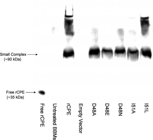
Clostridium perfringens enterotoxin (CPE) has a unique mechanism of action that results in the formation of large, sodium dodecyl sulfate-resistant complexes involving tight junction proteins; those complexes then induce plasma membrane permeability alterations in host intestinal epithelial cells, leading to cell death and epithelial desquamation. Previous deletion and point mutational studies mapped CPE receptor binding activity to the toxin's extreme C terminus. Those earlier analyses also determined that an N-terminal CPE region between residues D45 and G53 is required for large complex formation and cytotoxicity. To more finely map this N-terminal cytotoxicity region, site-directed mutagenesis was performed with recombinant CPE (rCPE). Alanine-scanning mutagenesis produced one rCPE variant, D48A, that failed to form large complexes or induce cytotoxicity, despite having normal ability to bind and form the small complex. Two saturation variants, D48E and D48N, also had a phenotype resembling that of the D48A variant, indicating that both size and charge are important at CPE residue 48. Another alanine substitution rCPE variant, I51A, was highly attenuated for large complex formation and cytotoxicity, but rCPE saturation variants I51L and I51V displayed a normal large complex formation and cytotoxicity phenotype. Collectively, these mutagenesis results identify a core CPE sequence extending from residues G47 to I51 that directly participates in large complex formation and cytotoxicity.
Figures
Similar articles
-
Identification of a Clostridium perfringens enterotoxin region required for large complex formation and cytotoxicity by random mutagenesis.Infect Immun. 1999 Nov;67(11):5634-41. doi: 10.1128/IAI.67.11.5634-5641.1999. Infect Immun. 1999. PMID: 10531210 Free PMC article.
-
Cysteine-scanning mutagenesis supports the importance of Clostridium perfringens enterotoxin amino acids 80 to 106 for membrane insertion and pore formation.Infect Immun. 2012 Dec;80(12):4078-88. doi: 10.1128/IAI.00069-12. Epub 2012 Sep 10. Infect Immun. 2012. PMID: 22966051 Free PMC article.
-
Identification of a prepore large-complex stage in the mechanism of action of Clostridium perfringens enterotoxin.Infect Immun. 2007 May;75(5):2381-90. doi: 10.1128/IAI.01737-06. Epub 2007 Feb 16. Infect Immun. 2007. PMID: 17307943 Free PMC article.
-
Clostridium perfringens enterotoxin acts by producing small molecule permeability alterations in plasma membranes.Toxicology. 1994 Feb 28;87(1-3):43-67. doi: 10.1016/0300-483x(94)90154-6. Toxicology. 1994. PMID: 8160188 Review.
-
An overview of Clostridium perfringens enterotoxin.Toxicon. 1996 Nov-Dec;34(11-12):1335-43. doi: 10.1016/s0041-0101(96)00101-8. Toxicon. 1996. PMID: 9027990 Review.
Cited by
-
Molecular characterization of podoviral bacteriophages virulent for Clostridium perfringens and their comparison with members of the Picovirinae.PLoS One. 2012;7(5):e38283. doi: 10.1371/journal.pone.0038283. Epub 2012 May 29. PLoS One. 2012. PMID: 22666499 Free PMC article.
-
The biology and pathogenicity of Clostridium perfringens type F: a common human enteropathogen with a new(ish) name.Microbiol Mol Biol Rev. 2024 Sep 26;88(3):e0014023. doi: 10.1128/mmbr.00140-23. Epub 2024 Jun 12. Microbiol Mol Biol Rev. 2024. PMID: 38864615 Free PMC article. Review.
-
The interaction of Clostridium perfringens enterotoxin with receptor claudins.Anaerobe. 2016 Oct;41:18-26. doi: 10.1016/j.anaerobe.2016.04.011. Epub 2016 Apr 16. Anaerobe. 2016. PMID: 27090847 Free PMC article. Review.
-
Potential Therapeutic Effects of Mepacrine against Clostridium perfringens Enterotoxin in a Mouse Model of Enterotoxemia.Infect Immun. 2019 Mar 25;87(4):e00670-18. doi: 10.1128/IAI.00670-18. Print 2019 Apr. Infect Immun. 2019. PMID: 30642896 Free PMC article.
-
Crystal structure of Clostridium perfringens enterotoxin displays features of beta-pore-forming toxins.J Biol Chem. 2011 Jun 3;286(22):19549-55. doi: 10.1074/jbc.M111.228478. Epub 2011 Apr 12. J Biol Chem. 2011. PMID: 21489981 Free PMC article.
References
-
- Abrahao, C., R. J. Carman, H. Hahn, and O. Liesenfeld. 2001. Similar frequency of detection of Clostridium perfringens enterotoxin and Clostridium difficile toxins in patients with antibiotic-associated diarrhea. Eur. J. Clin. Microbiol. Infect. Dis. 20:676-677. - PubMed
-
- Arnaout, M. A., S. L. Goodman, and J. P. Xiong. 2002. Coming to grips with integrin binding to ligands. Curr. Opin. Cell Biol. 14:641-651. - PubMed
Publication types
MeSH terms
Substances
Grants and funding
LinkOut - more resources
Full Text Sources
Research Materials
Miscellaneous
