. 2004 Dec;72(12):7084-95.
doi: 10.1128/IAI.72.12.7084-7095.2004.

A mutant of Mycobacterium tuberculosis H37Rv that lacks expression of antigen 85A is attenuated in mice but retains vaccinogenic potential

Affiliations

A mutant of Mycobacterium tuberculosis H37Rv that lacks expression of antigen 85A is attenuated in mice but retains vaccinogenic potential

Robert H Copenhaver et al. Infect Immun. 2004 Dec.

Abstract

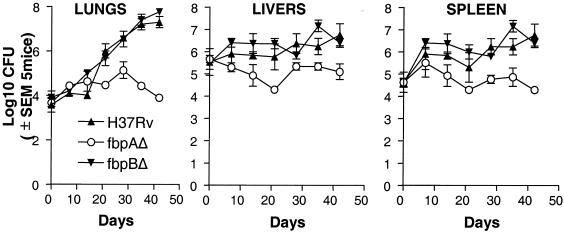
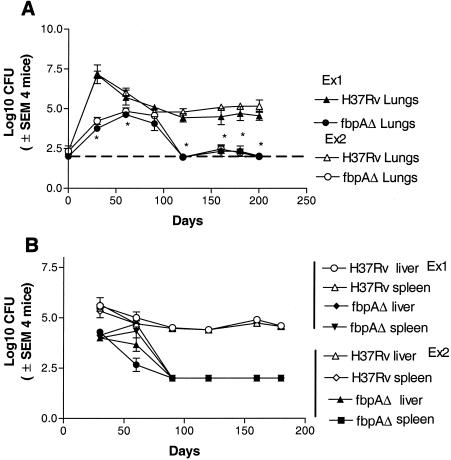
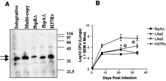
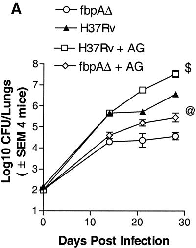
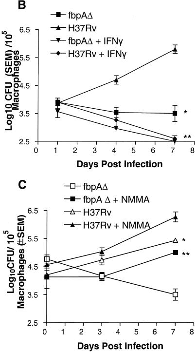
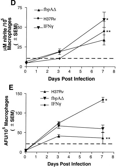
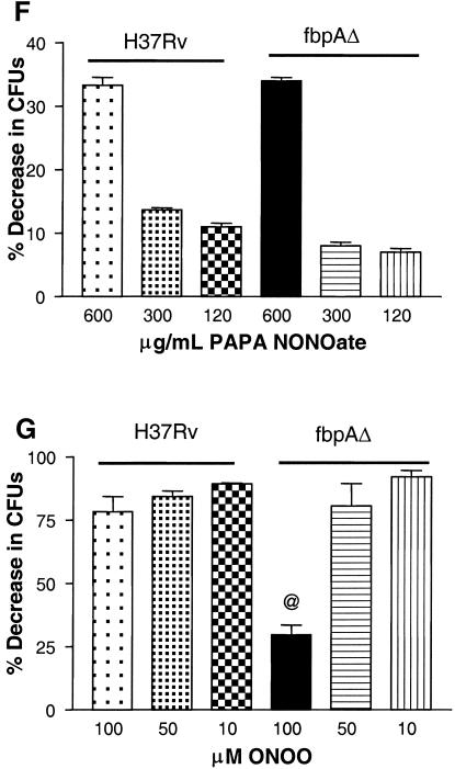
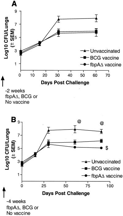
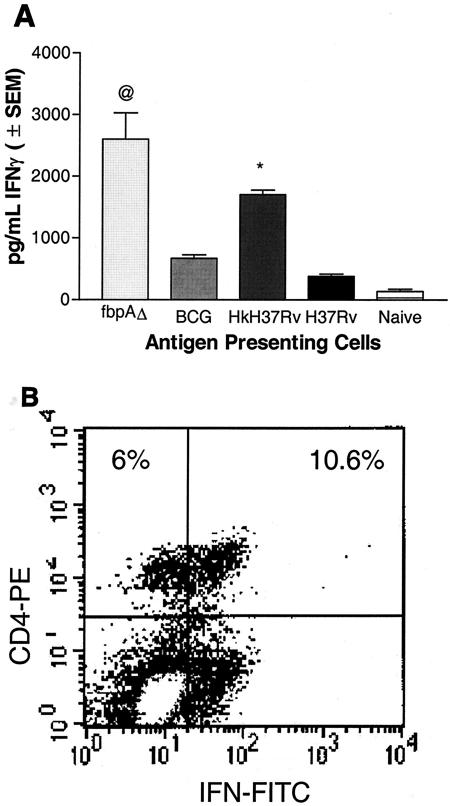
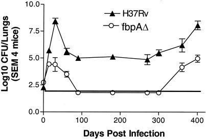
The fbpA and fbpB genes encoding the 85A and 85B proteins of Mycobacterium tuberculosis H37Rv, respectively, were disrupted, the mutants were examined for their ability to survive, and the strain lacking 85A (DeltafbpA) was tested for its ability to immunize mice. The DeltafbpA mutant was attenuated in mice after intravenous or aerosol infection, while replication of the DeltafbpB mutant was similar to that of the wild type. Complementation of the fbpA gene in DeltafbpA restored its ability to grow in the lungs of mice. The DeltafbpA mutant induced a stronger expression of pulmonary mRNA messages in mice for tumor necrosis factor alpha, interleukin-1 beta (IL-1beta), gamma interferon, IL-6, IL-2, and inducible nitric oxide (NO) synthase, which led to its decline, while H37Rv persisted despite strong immune responses. H37Rv and DeltafbpA both induced NO in macrophages and were equally susceptible to NO donors, although DeltafbpA was more susceptible in vitro to peroxynitrite and its growth was enhanced by NO inhibitors in mice and macrophages. Aerosol-infected mice, which cleared a low-dose DeltafbpA infection, resisted a challenge with virulent M. tuberculosis. Mice subcutaneously immunized with DeltafbpA or Mycobacterium bovis BCG and challenged with M. tuberculosis also showed similar levels of protection, marked by a reduction in the growth of challenged M. tuberculosis. The DeltafbpA mutant was thus attenuated, unlike DeltafbpB, but was also vaccinogenic against tuberculosis. Attenuation was incomplete, however, since DeltafbpA revived in normal mice after 370 days, suggesting that revival was due to immunosenescence but not compensation by the fbpB or fbpC gene. Antigen 85A thus affects susceptibility to peroxynitrite in M. tuberculosis and appears to be necessary for its optimal growth in mice.


Figures

FIG. 1.
FIG. 1.
Disruption of fibronectin binding protein A (fbpA) and fbpB genes has different effects on the growth profiles of M. tuberculosis H37Rv in mice. C57BL/6 mice were infected with an intravenous inoculum of 106 CFU of ΔfbpA (○), ΔfbpB (▾), or the wild type (▴) per mouse. Five mice per strain were sacrificed at the time points shown, and CFU counts were determined by plating organ homogenates on 7H11 agar. Both the wild type and the ΔfbpB mutant grow better in the lungs and moderately well in the livers and spleens. ΔfbpA grows initially but declines later in lungs, livers, and spleens. Its growth in lungs is markedly attenuated compared to both the wild type and the ΔfbpB mutant (P values were <0.001 for lung CFU and <0.007 for spleen and liver CFU on days 20 through 50 by the Mann-Whitney U test). SEM, standard error of the mean.
FIG. 2.
FIG. 2.
The ΔfbpA mutant is attenuated in mice after low-dose aerosol-induced infection. C57BL/6 mice were aerosol implanted with ΔfbpA or H37Rv, and CFU counts in lungs (A) and spleens and livers (B) were determined over time. Results of two independent experiments performed a year apart are shown, with four mice per strain per time point being sacrificed for CFU counts in each experiment. After an initial increase, CFU of ΔfbpA mutant in the lungs declined to low levels by day 90 postimplantation (A). H37Rv grows progressively until day 30 and then stabilizes over the next 150 days. ΔfbpA disseminates to spleens and livers, where it declines (B), while the wild type persists in spleens and livers in significant numbers. The broken horizontal line indicates the sensitivity of plating. SEM, standard error of the mean. (A) *, P value was <0.005 for ΔfbpA compared to H37Rv. (B) *, P value was <0.002 compared to H37Rv by the Mann-Whitney U test.
FIG. 3.
FIG. 3.
Effect of complementation of the fbpA gene in the ΔfbpA mutant. (A) The ΔfbpA mutant was complemented by using an integrative vector and multicopy plasmids. Western blot analysis of sodium dodecyl sulfate-polyacrylamide gel electrophoresis run on proteins isolated from H37Rv, ΔfbpA, and ΔfbpB mutants reveals that the ΔfbpA mutant lacks the antigen 85A protein band and the ΔfbpB mutant lacks the antigen 85B protein band. Use of multicopy plasmid transfectants shows restoration of the Ag85A protein, as does integrative vector for the Ag85A (fbpA) gene. The monoclonal antibody HYT27, which recognizes all three proteins of Ag85 complex, was used in the blots. Molecular weight standards (in thousands) are indicated on the left. Arrows indicate overlapping protein recognition. (B) C57BL/6 mice were aerosol implanted with wild-type H37Rv, ΔfbpA, and the two complemented strains (LAa2 integrative and LAa3 multicopy plasmid). At the indicated time points, four mice per strain were sacrificed and lung CFU was determined by plating on 7H11 agar. The growth of the ΔfbpA mutant is attenuated and similar to the profiles described above. Both of the complemented mutants grow better than the ΔfbpA mutant, although the integrative strain grows better than the strain with the multicopy plasmid. *, P value was <0.01 for LAa3 compared to ΔfbpA; @, P value was <0.01 day 21 and <0.008 on day 33 for LAa2 compared to ΔfbpA by the Mann-Whitney U test. Standard errors of the means (SEMs) for ΔfbpA are too low to be plotted.
FIG. 4.
FIG. 4.
Effect of nitric oxide (NO) on ΔfbpA growth in mice and macrophages. (A) Mice infected and treated with aminoguanidine (AG) show enhanced growth of ΔfbpA and of the wild type. @, P < 0.009; $, P < 0.008 (Mann-Whitney U test). (B and C) Naïve, IFN-γ-treated (400 U/ml) or NMMA-treated mouse macrophages were infected with ΔfbpA or H37Rv, and growth curves were plotted. (B) IFN-γ reduces baseline CFU. *, P value was <0.007 for ΔfbpA compared to H37Rv; **, P value was <0.02 for ΔfbpA compared to IFN-γ treatment and <0.003 for H37Rv compared to IFN-γ treatment (t test). (C) NMMA enhances growth of both strains. *, P value was <0.02 for ΔfbpA compared to NMMA treatment and <0.03 for H37Rv compared to NMMA treatment (t test). (D and E) Macrophages infected with ΔfbpA and H37Rv were measured for NO by using Griess reagent and ROS for 7 days postinfection with a DCFDA fluorescent probe. ΔfbpA and H37Rv induce similar levels of NO and ROS. (D) *, P value was <0.01 for IFN-γ compared to both strains; **, no significant difference found for ΔfbpA compared to H37Rv. (E) *, P value was <0.03 for IFN-γ compared to both strains; no significant difference was found for ΔfbpA compared to H37Rv. (F and G) Strains were treated with the NO donor, PAPA-NONOate, or peroxynitrite and measured for CFU counts, and results are expressed as the percent decrease from the vehicle control. ΔfbpA and H37Rv show nearly identical susceptibility to NO (F), but ΔfbpA is more susceptible to the 100 μM dose of peroxynitrite (G). @, P value was <0.01 (t test) for ΔfbpA compared to H37Rv. SEM, standard error of the mean.
FIG. 4.
FIG. 4.
Effect of nitric oxide (NO) on ΔfbpA growth in mice and macrophages. (A) Mice infected and treated with aminoguanidine (AG) show enhanced growth of ΔfbpA and of the wild type. @, P < 0.009; $, P < 0.008 (Mann-Whitney U test). (B and C) Naïve, IFN-γ-treated (400 U/ml) or NMMA-treated mouse macrophages were infected with ΔfbpA or H37Rv, and growth curves were plotted. (B) IFN-γ reduces baseline CFU. *, P value was <0.007 for ΔfbpA compared to H37Rv; **, P value was <0.02 for ΔfbpA compared to IFN-γ treatment and <0.003 for H37Rv compared to IFN-γ treatment (t test). (C) NMMA enhances growth of both strains. *, P value was <0.02 for ΔfbpA compared to NMMA treatment and <0.03 for H37Rv compared to NMMA treatment (t test). (D and E) Macrophages infected with ΔfbpA and H37Rv were measured for NO by using Griess reagent and ROS for 7 days postinfection with a DCFDA fluorescent probe. ΔfbpA and H37Rv induce similar levels of NO and ROS. (D) *, P value was <0.01 for IFN-γ compared to both strains; **, no significant difference found for ΔfbpA compared to H37Rv. (E) *, P value was <0.03 for IFN-γ compared to both strains; no significant difference was found for ΔfbpA compared to H37Rv. (F and G) Strains were treated with the NO donor, PAPA-NONOate, or peroxynitrite and measured for CFU counts, and results are expressed as the percent decrease from the vehicle control. ΔfbpA and H37Rv show nearly identical susceptibility to NO (F), but ΔfbpA is more susceptible to the 100 μM dose of peroxynitrite (G). @, P value was <0.01 (t test) for ΔfbpA compared to H37Rv. SEM, standard error of the mean.
FIG. 5.
FIG. 5.
Effect of prior infection with the ΔfbpA mutant on subsequent challenge with the Erdman strain. C57BL/6 mice were left uninfected or were aerosol infected with ΔfbpA. ΔfbpA reaches baseline levels by day 90. Mice (four per time point) were then challenged with the Erdman strain via aerosol. Mice previously infected with the ΔfbpA mutant reduce the growth of the challenge Erdman strain by more than 2 log10 (○) compared to untreated and challenged mice (•). SEM, standard error of the mean.
FIG. 6.
FIG. 6.
The ΔfbpA mutant is vaccinogenic against a challenge with the virulent Erdman strain in mice. C57BL/6 mice were left unimmunized (▴) or were immunized subcutaneously with two doses of live ΔfbpA (•) or BCG (Pasteur; ▪) 2 weeks apart, each at 106 CFU/mouse. Mice were challenged at either 2 (A) or 4 (B) weeks after the second dose of vaccine with an aerosol dose of the Erdman strain implanting into the lungs at ∼500 CFU/mouse. Four mice were sacrificed for each experiment at the time points shown, and lung CFU were determined. (A) Both BCG and ΔfbpA strains reduce the growth of the Erdman challenge strain at 2 weeks postvaccination. (B) ΔfbpA is more effective when the challenge is done at 4 weeks postvaccination. The Erdman strain grows better in naïve mice. $, P value was <0.01 for ΔfbpA compared to BCG (Mann-Whitney U test); @, P value was <0.009 for the Erdman strain compared to the ΔfbpA or BCG vaccine group (Mann-Whitney U test). SEM, standard error of the mean.
FIG. 7.
FIG. 7.
Immunogenicity of macrophages infected with H37Rv and ΔfbpA. (A) BM-derived macrophages from C57BL/6 mice were infected with ΔfbpA, BCG, live or heat-killed (Hk) H37Rv, or left uninfected. Splenic T cells from naïve mice or mice immunized with Hk-MTB were layered on macrophages, and the supernatants were tested for IFN-γ. ΔfbpA induces a stronger early IFN-γ response than BCG or live wild type. @, P value was <0.007 compared to BCG or H37Rv (t test). The positive control, Hk-H37Rv, also induced a higher response (P value was <0.009 compared to BCG or H37Rv). (B) Overlaid T cells were stained for phenotype and IFN-γ intrakine and analyzed by flow cytometry. ΔfbpA induces an early activation of IFN-γ-producing CD4+ T cells consistent with enhanced IFN-γ secretion (results shown are representative of three experiments). SEM, standard error of the mean.
FIG. 8.
FIG. 8.
Revival of wild-type H37Rv and the ΔfbpA mutant in mice. C57BL/6 mouse lungs were aerosol infected with ΔfbpA (○) and H37Rv (▴) (∼200 CFU/mouse). After an initial growth period, H37Rv stabilizes in lungs until day 300, after which organisms resume growth. Growth of the ΔfbpA mutant declines but resumes after day 350. SEM, standard error of the mean.

References

    1. Actor, J. K., T. Kuffner, C. S. Dezzutti, R. L. Hunter, and J. McNicholl. 1998. A flash-type bioluminescent immunoassay that is more sensitive than radioimaging: quantitative detection of cytokine cDNA in activated and resting human cells. J. Immunol. Methods 211:65-77. - PubMed
    1. Actor, J. K., M. Olsen, C. Jagannath, and R. L. Hunter. 1999. Relationship between survival, organism containment and granuloma formation in acute murine tuberculosis. J. Interferon Cytokine Res. 19:1183-1193. - PubMed
    1. Actor, J. K., E, Breij, H. Hoffmann, R. A. Wetsel, R. L. Hunter, and C. Jagannath. 2001. A role for complement C5 in organism containment and granulomatous response during murine tuberculosis in mice. Scand. J. Immunol. 53:464-474. - PubMed
    1. Armitige, L. Y., C. Jagannath, A. Wanger, and S. J. Norris. 2000. Disruption of the genes encoding antigen 85A and antigen 85B of Mycobacterium tuberculosis H37Rv: effect on growth in culture and in macrophages. Infect. Immun. 68:767-778. - PMC - PubMed
    1. Armitige, L. Y., S. J. Norris, A. Wanger, T. R. Sievert, and K. Takayama. Submitted for publication.

Publication types

MeSH terms

LinkOut - more resources