. 2004 Dec;72(12):7231-9.
doi: 10.1128/IAI.72.12.7231-7239.2004.

Differential regulation of inflammatory cytokine secretion by human dendritic cells upon Chlamydia trachomatis infection

Affiliations

Differential regulation of inflammatory cytokine secretion by human dendritic cells upon Chlamydia trachomatis infection

Ana Gervassi et al. Infect Immun. 2004 Dec.

Abstract

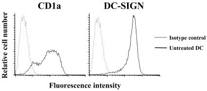
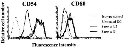
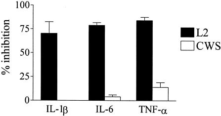
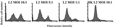
Chlamydia trachomatis is an obligate intracellular gram-negative bacterium responsible for a wide spectrum of diseases in humans. Both genital and ocular C. trachomatis infections are associated with tissue inflammation and pathology. Dendritic cells (DC) play an important role in both innate and adaptive immune responses to microbial pathogens and are a source of inflammatory cytokines. To determine the potential contribution of DC to the inflammatory process, human DC were infected with C. trachomatis serovar E or L2. Both C. trachomatis serovars were found to infect and replicate in DC. Upon infection, DC up-regulated the expression of costimulatory (B7-1) and cell adhesion (ICAM-1) molecules. Furthermore, chlamydial infection induced the secretion of interleukin-1beta (IL-1beta), IL-6, IL-8, IL-12p70, IL-18, and tumor necrosis factor alpha (TNF-alpha). The mechanisms involved in Chlamydia-induced IL-1beta and IL-18 secretion differed from those of the other cytokines. Chlamydia-induced IL-1beta and IL-18 secretion required infection with viable bacteria and was associated with the Chlamydia-induced activation of caspase-1 in infected host cells. In contrast, TNF-alpha and IL-6 secretion did not require that the Chlamydia be viable, suggesting that there are at least two mechanisms involved in the Chlamydia-induced cytokine secretion in DC. Interestingly, an antibody to Toll-like receptor 4 inhibited Chlamydia-induced IL-1beta, IL-6, and TNF-alpha secretion. The data herein demonstrate that DC can be infected by human C. trachomatis serovars and that chlamydial components regulate the secretion of various cytokines in DC. Collectively, these data suggest that DC play a role in the inflammatory processes caused by chlamydial infections.


Figures

FIG. 1.
FIG. 1.
Human monocyte-derived dendritic cell (DC) CD1a and DC-SIGN surface marker expression. DC were harvested after a 7-day incubation of human monocytes in GM-CSF- and IL-4-containing medium, and expression of CD1a and DC-SIGN was determined by fluorescence-activated cell sorter (FACS) analysis. Results are representative of four different donors.
FIG. 2.
FIG. 2.
Fluorescence microscopy (A and B) and TEM (C to E) of human monocyte-derived DC infected with C. trachomatis serovar E or L2. After human DC were infected with serovar E or L2 at an MOI of 10:1, DC were cultured for the indicated time periods and then fixed for fluorescence microscopy or TEM. (A) DC infected with serovar L2 for 24 h and stained with Chlamydia MOMP-specific antibody (green) and Evans blue (red). (B) DC infected with serovar E for 48 h and stained with Chlamydia MOMP-specific antibody (green) and Evans blue (red). (C and D) TEM of DC infected with serovar L2 for 24 h (C) and 48 h (D). (E) TEM of DC infected with serovar E for 72 h. Elementary bodies (EB) and reticulate bodies (RB) can be distinguished within the chlamydial inclusion.
FIG. 3.
FIG. 3.
Chlamydia infection up-regulates the expression of CD54 (ICAM-1) and CD80 (B7-1) on human monocyte-derived DC. DC were infected with C. trachomatis serovar L2 or E at a MOI of 10:1. Expression of cell surface markers CD54 and CD80 was determined by FACS analysis 24 h postinfection. Results are representative of four different donors.
FIG. 4.
FIG. 4.
TLR-dependent induction of IL-1β, IL-6, and TNF-α secretion by C. trachomatis in DC. DC (2 × 10⁵) from two human donors were treated with a neutralizing antibody to TLR4 (20 μg/ml) or isotype control antibody 2 h before and during stimulation. DC were either infected with C. trachomatis L2 (MOI, 1:1) or were stimulated with CWS from M. phlei (5 μg/ml). Cytokine levels were determined by ELISA in supernatants taken 24 h postinfection. Data are presented as percent inhibition of antibody treatment compared to the isotype control antibody results. Results are shown as means ± standard deviations (SD) for the two donors tested. The TLR4 antibody treatment used in the study was efficient at the reduction of IL-6 production in DC in response to LPS (1 ng/ml) from 3,600 pg/ml to 400 pg/ml. The figure is representative of three separate experiments.
FIG. 5.
FIG. 5.
Chloramphenicol inhibits IL-1β secretion but not IL-6 or TNF-α secretion in Chlamydia-infected DC. DC (2 × 10⁵) from two human donors were treated in the presence or absence of chloramphenicol (50 μg/ml) 2 h prior to and during infection with C. trachomatis serovar L2 (MOI, 10:1). Cytokine levels were determined by ELISA in supernatants taken 24 h postinfection. Data represent means ± SD of cytokine release from two human donors. The figure is representative of three separate experiments.
FIG. 6.
FIG. 6.
Both viable and inactivated Chlamydia bacteria induce the production of pro-IL-1β by human monocyte-derived DC. DC (1.5 × 10⁶) were infected with C. trachomatis serovar L2 (MOI, 10:1) (lane 2), pulsed with heat-inactivated serovar L2 (MOI, 10:1) (lane 3), or treated with 1 μg of LPS/ml (lane 4) as indicated. Untreated DC (lane 1) and mature recombinant IL-1β (lane 5) were used as controls. DC lysates were prepared 24 h after treatment, and the presence of IL-1β in DC lysates was analyzed by Western blotting. The results are representative of three independent experiments.
FIG. 7.
FIG. 7.
Active caspase-1 is detected in human monocyte-derived DC after infection with viable but not heat-killed C. trachomatis. DC were either infected with C. trachomatis serovar L2 (L2) at MOIs of 10, 5, and 1:1 or pulsed with heat inactivated serovar L2 (HK L2) (MOI, 10:1). Intracellular levels of active caspase-1 were determined by FACS analysis in DC 24 h postinfection. Results of one representative experiment of five are shown.
FIG. 8.
FIG. 8.
Infection of DC with viable C. trachomatis but not culture with heat-inactivated C. trachomatis induces the production of IL-18. Monocyte-derived DC (2 × 10⁵) were either infected as indicated with C. trachomatis serovar L2 (L2) and serovar E (E), pulsed with heat-inactivated L2 (HK L2) and heat-inactivated E (HK E) at an MOI of 10:1, or left untreated (Medium). IL-1β and IL-18 levels were determined by ELISA in supernatants taken 24 h posttreatment. Means ± SD were calculated for two different donors. The results are representative of three independent experiments.

References

    1. Abu el-Asrar, A. M., K. Geboes, K. F. Tabbara, S. A. al Kharashi, L. Missotten, and V. Desmet. 1998. Immunopathogenesis of conjunctival scarring in trachoma. Eye 12(Pt. 3a):453-460. - PubMed
    1. Antonopoulos, C., M. Cumberbatch, R. J. Dearman, R. J. Daniel, I. Kimber, and R. W. Groves. 2001. Functional caspase-1 is required for Langerhans cell migration and optimal contact sensitization in mice. J. Immunol. 166:3672-3677. - PubMed
    1. Azuma, I., E. E. Ribi, T. J. Meyer, and B. Zbar. 1974. Biologically active components from mycobacterial cell walls. I. Isolation and composition of cell wall skeleton and component P3. J. Natl. Cancer Inst. 52:95-101. - PubMed
    1. Babcock, T. A., and J. M. Carlin. 2000. Transcriptional activation of indoleamine dioxygenase by interleukin 1 and tumor necrosis factor alpha in interferon-treated epithelial cells. Cytokine 12:588-594. - PubMed
    1. Bhardwaj, N. 2001. Processing and presentation of antigens by dendritic cells: implications for vaccines. Trends Mol. Med. 7:388-394. - PubMed

Publication types