. 2004 Dec;72(12):7294-305.
doi: 10.1128/IAI.72.12.7294-7305.2004.

Mannose-resistant Proteus-like fimbriae are produced by most Proteus mirabilis strains infecting the urinary tract, dictate the in vivo localization of bacteria, and contribute to biofilm formation

Affiliations

Mannose-resistant Proteus-like fimbriae are produced by most Proteus mirabilis strains infecting the urinary tract, dictate the in vivo localization of bacteria, and contribute to biofilm formation

Angela M Jansen et al. Infect Immun. 2004 Dec.

Abstract

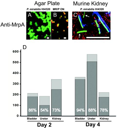
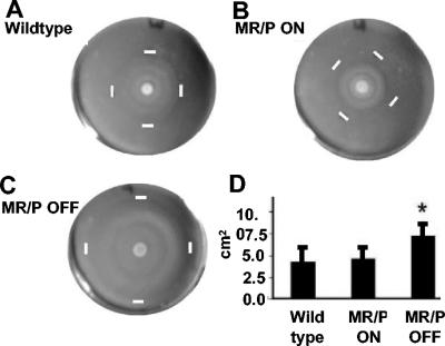
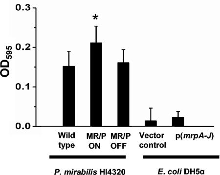
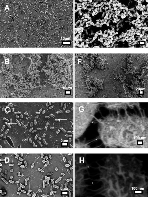
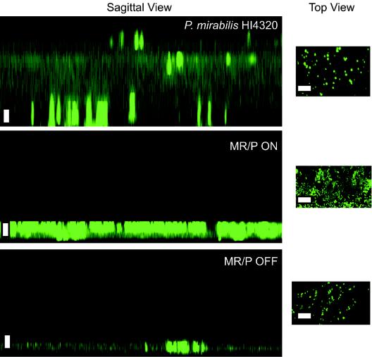
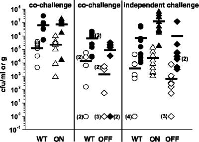
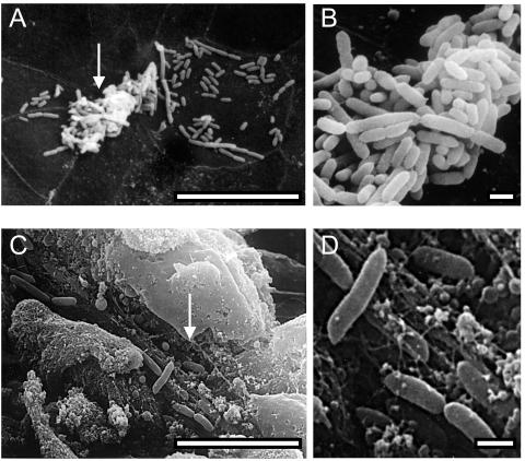
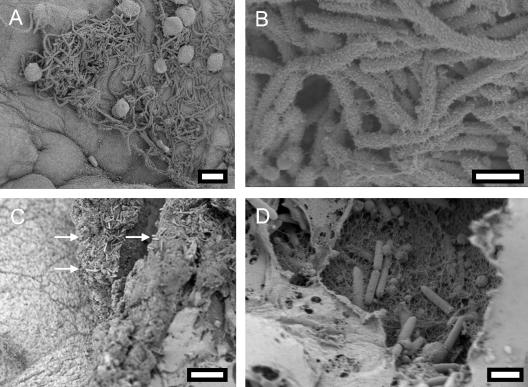
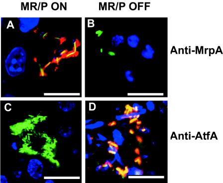
Proteus mirabilis, an etiologic agent of complicated urinary tract infections, expresses mannose-resistant Proteus-like (MR/P) fimbriae whose expression is phase variable. Here we examine the role of these fimbriae in biofilm formation and colonization of the urinary tract. The majority of wild-type P. mirabilis cells in transurethrally infected mice produced MR/P fimbriae. Mutants that were phase-locked for either constitutive expression (MR/P ON) or the inability to express MR/P fimbriae (MR/P OFF) were phenotypically distinct and swarmed at different rates. The number of P. mirabilis cells adhering to bladder tissue did not appear to be affected by MR/P fimbriation. However, the pattern of adherence to the bladder surface was strikingly different. MR/P OFF colonized the lamina propria underlying exfoliated uroepithelium, while MR/P ON colonized the luminal surfaces of bladder umbrella cells and not the exfoliated regions. Wild-type P. mirabilis was usually found colonizing intact uroepithelium, but it occasionally adhered to exfoliated areas. MR/P ON formed significantly more biofilm than either P. mirabilis HI4320 (P = 0.03) or MR/P OFF (P = 0.05). MR/P OFF was able to form a biofilm similar to that of the wild type. MR/P ON formed a three-dimensional biofilm structure as early as 18 h after the initiation of the biofilm, while MR/P OFF and the wild type did not. After 7 days, however, P. mirabilis HI4320 formed a 65-mum-thick biofilm, while the thickest MR/P ON and MR/P OFF biofilms were only 12 mum thick. We concluded that MR/P fimbriae are expressed by most P. mirabilis cells infecting the urinary tract, dictate the localization of bacteria in the bladder, and contribute to biofilm formation.


Figures

FIG. 1.
FIG. 1.
Expression of MR/P fimbriae in experimentally infected murine urinary tract. (A) An overnight culture of P. mirabilis HI4320(pBAC001) was spread onto nonswarming LB agar plates and incubated overnight at 37°C in 5% CO2. Colonies were washed from the plates with PBS, and bacteria were diluted to a concentration of 109 bacteria/ml. P. mirabilis HI4320(pBAC001) was spread onto a glass slide and incubated with anti-MrpA for 1 h at 37°C. After three 6-min washes, a goat anti-rabbit antibody conjugated to Alexa Fluor 568 was used to visualize MrpA (red). (B) MR/P ON(pBAC001) bacteria were treated in the same manner as a positive control for MR/P production. (C) Female CBA mice were inoculated with 50 μl of the P. mirabilis HI4320(pBAC001) bacterial suspension as described in Materials and Methods. Bacteria in tissue sections (10 μm) were stained with anti-MrpA (blue) and anti-actin (red) antibodies. Six bacteria produced MR/P fimbriae (blue and green), while two bacteria did not produce MR/P fimbriae (green only, arrows). Bars = 1 μm (A and B) and 10 μm (C). (D) Tissues from the experimentally infected mice were used to quantify the percentages of P. mirabilis HI4320(pBAC001) bacteria expressing MR/P fimbriae (percentages are shown in the dark gray portion of each bar). The y axis depicts the total number of bacteria counted.
FIG. 2.
FIG. 2.
MR/P OFF swarms faster than MR/P ON or wild-type P. mirabilis. Swarming plates for wild-type P. mirabilis HI4320 (A), MR/P ON (B), and MR/P OFF (C) are shown. Bacteria were spotted onto the center of the agar surface, and the plates were incubated at 37°C. Measurements were taken after 12.5 h. White bars indicate the outer limits of the swarm. (D) The average area of each swarm was determined by using the formula for the area of an ellipse [area = (height × width × π)/4]. Areas were determined for nine swarm zones for each strain. *, P < 0.0028.
FIG. 3.
FIG. 3.
MR/P fimbriae increase biofilm production in P. mirabilis. Aliquots of overnight cultures of wild-type P. mirabilis HI4320, MR/P ON, MR/P OFF, E. coli DH5α (vector control), and DH5α(mrpA-J) were standardized to an OD600 of 0.5 and diluted 1:10 in fresh sterile urine in 96-well plates. After 48 h at 30°C, with decanting of the medium and planktonic bacteria at 24 h, the biofilms were washed with sterile distilled water and a crystal violet biofilm assay was performed as described in Materials and Methods. Data are the averages of quadruplicate experiments that were performed three times. *, P < 0.01 for MR/P ON compared to the wild type and MR/P OFF.
FIG. 4.
FIG. 4.
SEM of 18-h biofilm grown on glass. Aliquots of overnight cultures of P. mirabilis HI4320, MR/P ON, and MR/P OFF were standardized to an OD600 of 0.05 and diluted 1:10 in fresh sterile urine on cover glass. The biofilms were incubated for 18 h at 30°C. The biofilms were rinsed with copious amounts of sterile distilled water and then processed for SEM as described in Materials and Methods. (A and D) MR/P OFF; (B and E to H) MR/P ON (asterisks denote surface structures that are consistent in size and shape with fimbriae); (C) wild-type P. mirabilis HI4320.
FIG. 5.
FIG. 5.
Overhead (xy) and sagittal (xz) images of wild-type and MR/P phase-locked mutants. All strains were transformed with pBAC001 (a plasmid encoding GFP) and grown at 30°C in urine placed on a cover glass. The cultures were grown in urine with ampicillin (100 μg/ml) for 7 days without shaking. Saturated urine was replaced with fresh urine and ampicillin every 48 h. After 7 days, each biofilm was rinsed to remove nonadherent bacteria, and the remaining attached cells were examined via confocal microscopy with a 63× oil immersion objective (numerical aperture, 1.4) on a Zeiss LSM410 instrument. Sagittal images were created from a collection of 30 consecutive z-series scans of the wild-type and mutant biofilms. The step size between each z section was 2 μm. Sagittal images were generated with the LSM410 PC software package. Bar = 10 μm.
FIG. 6.
FIG. 6.
Numbers of adherent and nonadherent P. mirabilis cells remaining in murine bladders after washing. Strains were tested in cochallenge experiments (eight mice per group) with a transurethrally inoculated, ∼108-CFU equal mixture of wild-type P. mirabilis HI4320 and MR/P ON or wild-type P. mirabilis HI4320 and MR/P OFF. Independent challenges (10 mice/group) were also conducted with each strain. Bladders were washed at 24 h postinoculation and then homogenized. Both the wash fluid and the homogenized bladders were immediately plated to determine the numbers of nonadherent bacteria (open symbols) present in the wash fluid and the numbers of adherent bacteria (closed symbols) present in the homogenized bladders. Bars represent the means for all mice in each group. All values examined by the nonparametric Mann-Whitney test showed no significant differences among the strains' abilities to adhere in the bladder.
FIG. 7.
FIG. 7.
SEM micrographs of wild-type P. mirabilis HI4320 colonizing the murine bladder. Mice were transurethrally inoculated with 108 CFU of wild-type P. mirabilis HI4320. At 7 days postinoculation, the ureters of mice infected with wild-type P. mirabilis were ligated with surgical thread, the bladders were infused through the urethra with fixative (2% glutaraldehyde, 2% formaldehyde), and then the urethras were tied off. Bladders were aseptically removed and processed for SEM as described in Materials and Methods. Wild-type P. mirabilis HI4320 colonized the bladder uroepithelium (A and B) and, occasionally, areas of exfoliated bladder cells (C and D). Arrows point to areas that are enlarged to the right. Bars = 10 μm (A and C) and 1 μm (B and D).
FIG. 8.
FIG. 8.
SEM micrographs of MR/P ON and MR/P OFF cells colonizing the murine bladder. Mice were transurethrally inoculated with 108 CFU of the mutants in independent experiments. At 4 days postinoculation, the ureters of infected mice were ligated with surgical thread, and the bladders were infused through the urethra with fixative (2% glutaraldehyde, 2% formaldehyde). After the urethras were tied off, the bladders were aseptically removed and processed for SEM as described in Materials and Methods. MR/P ON colonized the bladder uroepithelium (A and B), while MR/P OFF colonized the lamina propria where bladder cells had sloughed off (C [arrows point to MR/P OFF] and D). Bars = 10 μm (A), 2 μm (B and D), and 15 μm (C).
FIG. 9.
FIG. 9.
MR/P and ATF expression during ascending UTI. Representative confocal laser scanning micrographs show that the phase-locked mutants continued to either express MR/P fimbriae (MR/P ON) (A) or to not express MR/P fimbriae (MR/P OFF) (B) during experimental UTI. Mice were infected transurethrally with either MR/P ON(pBAC001) or MR/P OFF(pBAC001). After 2 days, the mice were euthanized and their bladders were removed and processed for confocal microscopy as described in Materials and Methods. Sections (10 μm) were stained with a 1:200 dilution of rabbit anti-MrpA, followed by staining with goat anti-rabbit IgG conjugated to Alexa Fluor 568. Tissue sections were mounted with Vectashield containing DAPI. (A) MR/P ON-infected tissue. Green, GFP-expressing P. mirabilis; blue, DAPI-stained bladder cell nuclei; red, rabbit anti-MrpA reacted with goat anti-rabbit IgG conjugated to Alexa Fluor 568; yellow, colocalization of bacteria and fimbriae. (B) MR/P OFF-infected tissue. Blue, DAPI-stained bladder nuclei; green, GFP-expressing P. mirabilis. (C) MR/P ON does not express ATF (green bacteria). (D) MR/P OFF expresses ATF (green bacteria colocalizing with red-stained ATF appear yellow). Sections (10 μm) were stained with a 1:500 dilution of a rabbit anti-ATF serum, followed by staining with goat anti-rabbit IgG conjugated to Alexa Fluor 568. Tissue sections were mounted with Vectashield containing DAPI. For MR/P ON-infected tissue: green, GFP-expressing P. mirabilis; blue, DAPI-stained bladder cell nuclei. For MR/P OFF-infected tissue: green, GFP-expressing P. mirabilis; red, rabbit anti-ATF serum reacted with goat anti-rabbit IgG conjugated to Alexa Fluor 568; blue, DAPI-stained bladder nuclei (D). Bars = 10 μm.

References

    1. Abraham, J. M., C. S. Freitag, J. R. Clements, and B. I. Eisenstein. 1985. An invertible element of DNA controls phase variation of type 1 fimbriae of Escherichia coli. Proc. Natl. Acad. Sci. USA 82:5724-5727. - PMC - PubMed
    1. Anderson, G. G., J. J. Palermo, J. D. Schilling, R. Roth, J. Heuser, and S. J. Hultgren. 2003. Intracellular bacterial biofilm-like pods in urinary tract infections. Science 301:105-107. - PubMed
    1. Bahrani, F. K., G. Massad, C. V. Lockatell, D. E. Johnson, R. G. Russell, J. W. Warren, and H. L. Mobley. 1994. Construction of an MR/P fimbrial mutant of Proteus mirabilis: role in virulence in a mouse model of ascending urinary tract infection. Infect. Immun. 62:3363-3371. - PMC - PubMed
    1. Bahrani, F. K., and H. L. Mobley. 1993. Proteus mirabilis MR/P fimbriae: molecular cloning, expression, and nucleotide sequence of the major fimbrial subunit gene. J. Bacteriol. 175:457-464. - PMC - PubMed
    1. Belas, R. 1994. Expression of multiple flagellin-encoding genes of Proteus mirabilis. J. Bacteriol. 176:7169-7181. - PMC - PubMed

Publication types