

doi: 10.1128/IAI.72.12.7311-7314.2004.
Expression of a beta-defensin mRNA, lingual antimicrobial peptide, in bovine mammary epithelial tissue is induced by mastitis
Affiliations
- PMID: 15557657
- PMCID: PMC529112
- DOI: 10.1128/IAI.72.12.7311-7314.2004
Item in Clipboard
Expression of a beta-defensin mRNA, lingual antimicrobial peptide, in bovine mammary epithelial tissue is induced by mastitis
Infect Immun.
2004 Dec.
Abstract
The expression of a beta-defensin, the lingual antimicrobial peptide (LAP), in response to mastitis was investigated by real-time PCR of RNA from mastitic and control udder quarters. There was a positive relationship between somatic cell count in milk and LAP expression. In situ hybridization showed that LAP mRNA was expressed in epithelial cells of mastitic tissue. These results suggest that LAP plays a role in the innate immune response to mastitis.
Figures
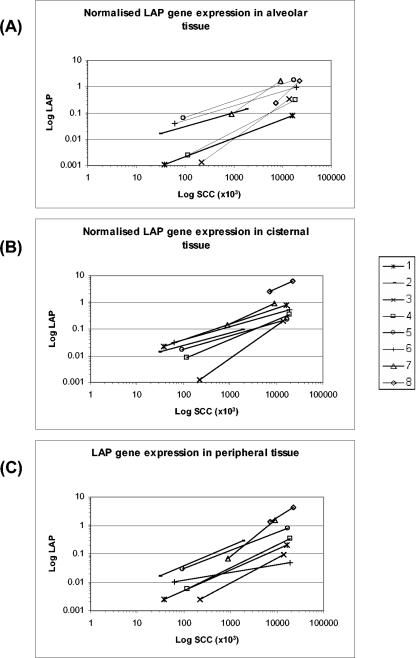
Levels of LAP gene expression in three regions of the bovine mammary gland in each of the eight cows. Normalized amounts of LAP mRNA in individual udder quarters for all cows are plotted. Each point is represented by a symbol with lines connecting noninfected and infected tissue samples from each cow. The slope of the line represents the correlation between log LAP gene expression and log SCCs. (A) Alveolar tissue; (B) cisternal tissue; (C) peripheral tissue.
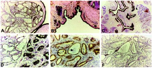
In situ hybridization of the LAP 35S-labeled probe to infected tissue sections of the bovine mammary gland. The tissue sections were probed with the 35S-labeled LAP sense and antisense probes. Hybridized sections were exposed for 40 days under nuclear emulsion at 4°C. High-intensity expression is demonstrated as dense black labeling by silver grains over the cells. (A) Cow 3, infected cisternal tissue. Magnification, ×100. (B) Cow 2, infected teat epithelium. Magnification, ×100. (C) Cow 2, subclinical infection (low SCC) cisternal tissue. Magnification, ×40. (D) Cow 3, infected cisternal-alveolar junction. Magnification, ×200. (E) Cow 3, subclinical alveolar tissue. No signal is detected in the white blood cells (arrow) within the alveoli. (F) Control serial tissue of the sample from panel E probed with the LAP sense probe. Magnification, ×200.
References
-
- Cullor, J., S. Wood, W. Smith, L. Panico, and M. Selsted. 1991. Bactericidal potency and mechanistic specificity of neutrophil defensins against bovine mastitis pathogens. Vet. Microbiol. 29:49-58. - PubMed
-
- Harder, J., J. Bartels, E. Christophers, and J. M. Schroder. 1997. A peptide antibiotic from human skin. Nature 387:861. - PubMed
-
- Hill, A. W., A. L. Shears, and K. G. Hibbitt. 1978. The elimination of serum-resistant Escherichia coli from experimentally infected single mammary glands of healthy cows. Res. Vet. Sci. 25:89-93. - PubMed
-
- Jia, H. P., T. Starner, M. Ackermann, P. Kirby, B. F. Tack, and P. B. McCray, Jr. 2001. Abundant human beta-defensin-1 expression in milk and mammary gland epithelium. J. Pediatr. 138:109-112. - PubMed
Publication types
MeSH terms
Substances
LinkOut - more resources
Full Text Sources
Research Materials
