

Microvascular density and hypoxia-inducible factor pathway in pancreatic endocrine tumours: negative correlation of microvascular density and VEGF expression with tumour progression
- PMID: 15558070
- PMCID: PMC2361752
- DOI: 10.1038/sj.bjc.6602245
Microvascular density and hypoxia-inducible factor pathway in pancreatic endocrine tumours: negative correlation of microvascular density and VEGF expression with tumour progression
Abstract
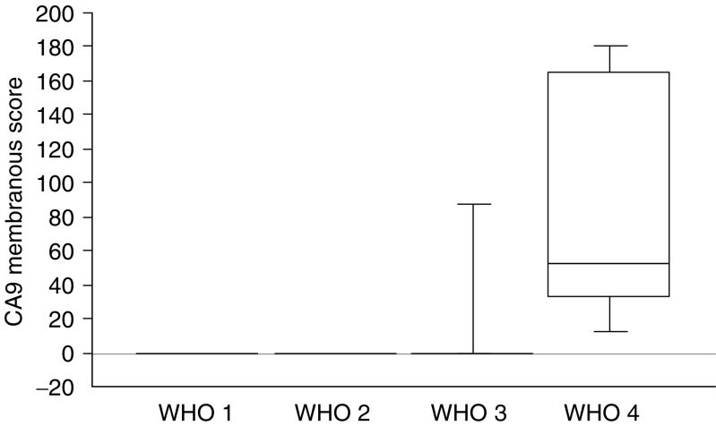
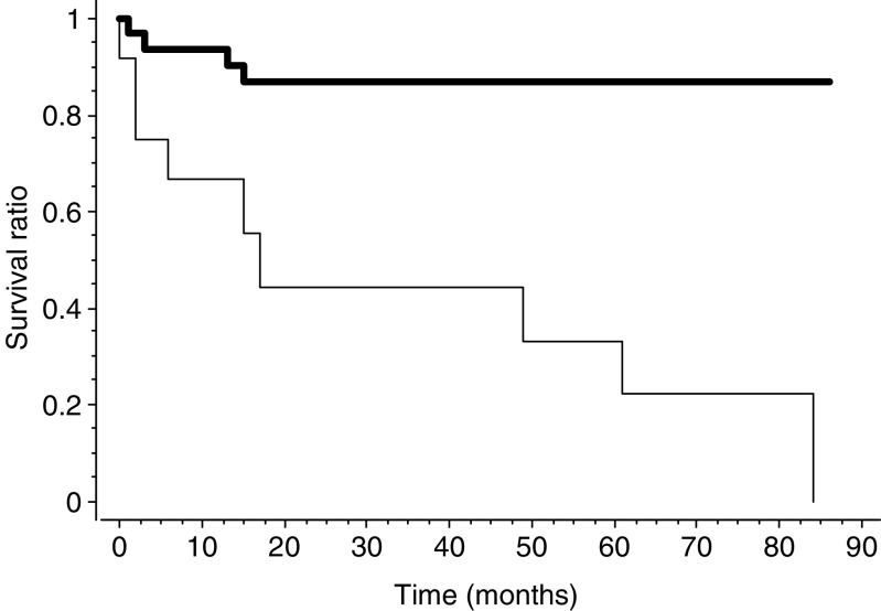
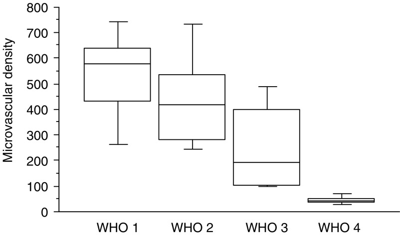
Tumour-associated angiogenesis is partly regulated by the hypoxia-inducible factor (HIF) pathway. Endocrine tumours are highly vascularised and the molecular mechanisms of their angiogenesis are not fully delineated. The aim of this study is to evaluate angiogenesis and expression of HIF-related molecules in a series of patients with pancreatic endocrine tumours (PETs). The expression of vascular endothelial growth factor (VEGF), HIF-1alpha, HIF-2alpha and carbonic anhydrase 9 (CA9) was examined by immunohistochemistry in 45 patients with PETs and compared to microvascular density (MVD), endothelial proliferation, tumour stage and survival. Microvascular density was very high in PETs and associated with a low endothelial index of proliferation. Microvascular density was significantly higher in benign PETs than in PETs of uncertain prognosis, well-differentiated and poorly differentiated carcinomas (mean values: 535, 436, 252 and 45 vessels mm(-2), respectively, P < 0.0001). Well-differentiated tumours had high cytoplasmic VEGF and HIF-1alpha expression. Poorly differentiated carcinomas were associated with nuclear HIF-1alpha and membranous CA9 expression. Low MVD (P = 0.0001) and membranous CA9 expression (P = 0.0004) were associated with a poorer survival. Contrary to other types of cancer, PETs are highly vascularised, but poorly angiogenic tumours. As they progress, VEGF expression is lost and MVD significantly decreases. The regulation of HIF signalling appears to be specific in pancreatic endocrine tumours.
Figures


References
-
- Bergers G, Benjamin LE (2003) Tumorigenesis and the angiogenic switch. Nat Rev Cancer 3: 401–410 - PubMed
-
- Bernini GP, Moretti A, Bonadio AG, Menicagli M, Viacava P, Naccarato AG, Iacconi P, Miccoli P, Salvetti A (2002) Angiogenesis in human normal and pathologic adrenal cortex. J Clin Endocrinol Metab 87: 4961–4965 - PubMed
-
- Birner P, Schindl M, Obermair A, Plank C, Breitenecker G, Oberhuber G (2000) Overexpression of hypoxia-inducible factor 1alpha is a marker for an unfavorable prognosis in early-stage invasive cervical cancer. Cancer Res 60: 4693–4696 - PubMed
-
- Blouw B, Song H, Tihan T, Bosze J, Ferrara N, Gerber HP, Johnson RS, Bergers G (2003) The hypoxic response of tumors is dependent on their microenvironment. Cancer Cell 4: 133–146 - PubMed
-
- Carmeliet P (2003) Angiogenesis in health and disease. Nat Med 9: 653–660 - PubMed
MeSH terms
Substances
LinkOut - more resources
Full Text Sources
Other Literature Sources
Medical
Research Materials
