

New views on retinal axon development: a navigation guide
- PMID: 15558486
- PMCID: PMC3683942
- DOI: 10.1387/ijdb.041899fm
New views on retinal axon development: a navigation guide
Abstract
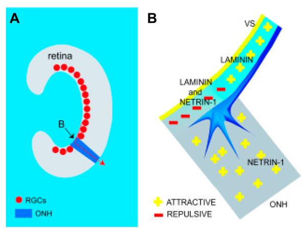
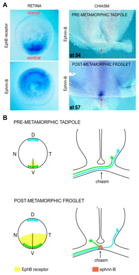
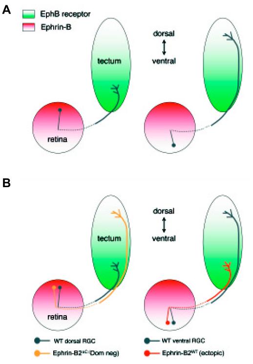
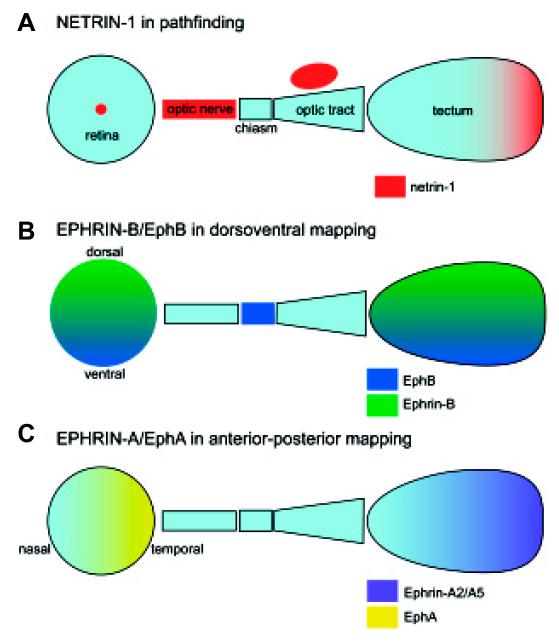
The eye is a peripheral outpost of the central nervous system (CNS) where the retinal ganglion cells (RGCs) reside. RGC axons navigate to their targets in a remarkably stereotyped and error-free manner and it is this process of directed growth that underlies the complex organization of the adult brain. The RGCs are the only retinal neurons to project into the brain and their peripheral location makes them an unusually accessible population of projection neurons for experiments involving in vivo gene transfer, anatomical tracing, transplantation and in vitro culture. In this paper, we review recent findings that have contributed to our understanding of some of the guidance decisions that axons make in the developing visual system. We look at two choice points in the pathway, the optic nerve head (onh) and the midline chiasm, and discuss evidence that supports the idea that key molecules in guiding axon growth at these junctures are netrin-1 (onh) and ephrin-B (chiasm). In the optic tectum where RGC axon terminals are arrayed in topographic order, we present experimental evidence to suggest that in the dorso-ventral dimension, the B-type ephrins and Eph receptors are of prime importance, possibly through attractive interactions. This complements the anterior-posterior topographic mapping known to be mediated through A-type ephrin/Eph repulsive interactions. An emerging theme is that guidance molecules such as ephrin-B and netrin-1 have complex patterns of restricted expression in the pathway and play multiple and changing roles in axon guidance.
Figures
Similar articles
-
Kinase independent function of EphB receptors in retinal axon pathfinding to the optic disc from dorsal but not ventral retina.Development. 2000 Mar;127(6):1231-41. doi: 10.1242/dev.127.6.1231. Development. 2000. PMID: 10683176
-
Altered midline axon pathways and ectopic neurons in the developing hypothalamus of netrin-1- and DCC-deficient mice.J Neurosci. 1999 Nov 15;19(22):9900-12. doi: 10.1523/JNEUROSCI.19-22-09900.1999. J Neurosci. 1999. PMID: 10559399 Free PMC article.
-
Heparan sulfate regulates intraretinal axon pathfinding by retinal ganglion cells.Invest Ophthalmol Vis Sci. 2011 Aug 22;52(9):6671-9. doi: 10.1167/iovs.11-7559. Invest Ophthalmol Vis Sci. 2011. PMID: 21743013 Free PMC article.
-
Ganglion cell axon pathfinding in the retina and optic nerve.Semin Cell Dev Biol. 2004 Feb;15(1):125-36. doi: 10.1016/j.semcdb.2003.09.006. Semin Cell Dev Biol. 2004. PMID: 15036215 Review.
-
Molecular mechanisms of optic axon guidance.Naturwissenschaften. 2005 Dec;92(12):549-61. doi: 10.1007/s00114-005-0042-5. Epub 2005 Oct 12. Naturwissenschaften. 2005. PMID: 16220285 Review.
Cited by
-
Role of Netrin-1 Signaling in Nerve Regeneration.Int J Mol Sci. 2017 Feb 24;18(3):491. doi: 10.3390/ijms18030491. Int J Mol Sci. 2017. PMID: 28245592 Free PMC article. Review.
-
Netrin participates in the development of retinotectal synaptic connectivity by modulating axon arborization and synapse formation in the developing brain.J Neurosci. 2009 Sep 9;29(36):11065-77. doi: 10.1523/JNEUROSCI.0947-09.2009. J Neurosci. 2009. PMID: 19741113 Free PMC article.
-
Nitric oxide as a putative retinal axon pathfinding and target recognition cue in Xenopus laevis.Impulse (Columbia). 2011 Jan 1;2010:1-12. Impulse (Columbia). 2011. PMID: 21847432 Free PMC article.
-
Classification of genes and putative biomarker identification using distribution metrics on expression profiles.PLoS One. 2010 Feb 4;5(2):e9056. doi: 10.1371/journal.pone.0009056. PLoS One. 2010. PMID: 20140228 Free PMC article.
-
Retinal regeneration is facilitated by the presence of surviving neurons.Dev Neurobiol. 2014 Sep;74(9):851-76. doi: 10.1002/dneu.22167. Epub 2014 Feb 18. Dev Neurobiol. 2014. PMID: 24488694 Free PMC article.
References
-
- ANDERSON RB, HOLT CE. Expression of unc-5 in the developing Xenopus visual system. Mech Dev. 2002;118:157–60. - PubMed
-
- BIRGBAUER E, COWAN CA, SRETAVAN DW, HENKEMEYER M. Kinase independent function of ephb receptors in retinal axon pathfinding to the optic disc from dorsal but not ventral retina. Development. 2000;127:1231–41. - PubMed
-
- BIRGBAUER E, OSTER SF, SEVERIN CG, SRETAVAN DW. Retinal axon growth cones respond to ephb extracellular domains as inhibitory axon guidance cues. Development. 2001;128:3041–8. - PubMed
-
- BRAISTED JE, MCLAUGHLIN T, WANG HU, FRIEDMAN GC, ANDERSON DJ, O’LEARY DD. Graded and lamina-specific distributions of ligands of ephb receptor tyrosine kinases in the developing retinotectal system. Dev Biol. 1997;191:14–28. - PubMed
-
- BRENNAN C, MONSCHAU B, LINDBERG R, GUTHRIE B, DRESCHER U, BONHOEFFER F, HOLDER N. Two eph receptor tyrosine kinase ligands control axon growth and may be involved in the creation of the retinotectal map in the zebrafish. Development. 1997;124:655–64. - PubMed
Publication types
MeSH terms
Substances
Grants and funding
LinkOut - more resources
Full Text Sources
Miscellaneous
