. 2005 Jan;76(1):112-27.
doi: 10.1086/427268. Epub 2004 Nov 22.

Extreme heterogeneity in the molecular events leading to the establishment of chiasmata during meiosis i in human oocytes

Affiliations

Extreme heterogeneity in the molecular events leading to the establishment of chiasmata during meiosis i in human oocytes

Michelle L Lenzi et al. Am J Hum Genet. 2005 Jan.

Abstract

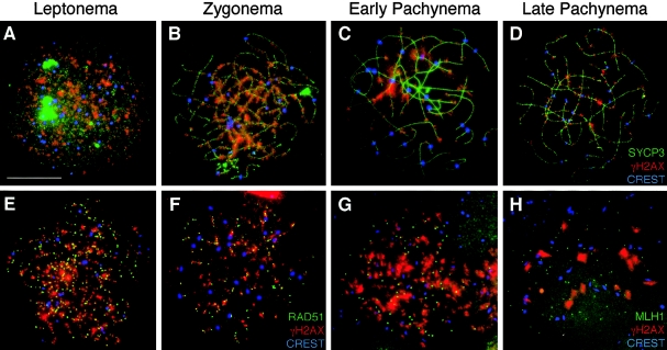
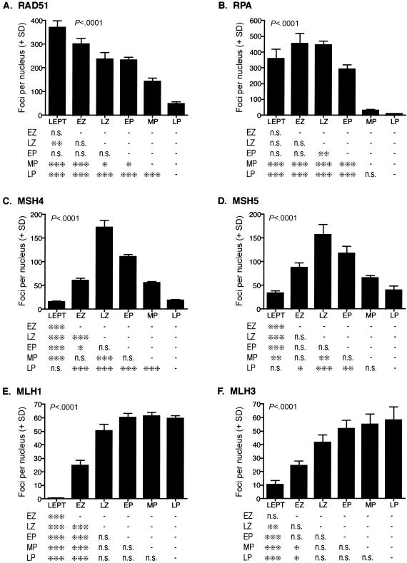
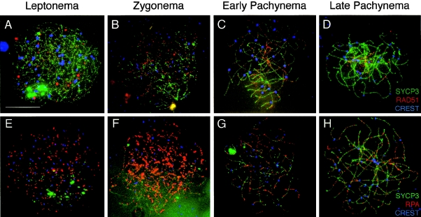
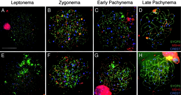
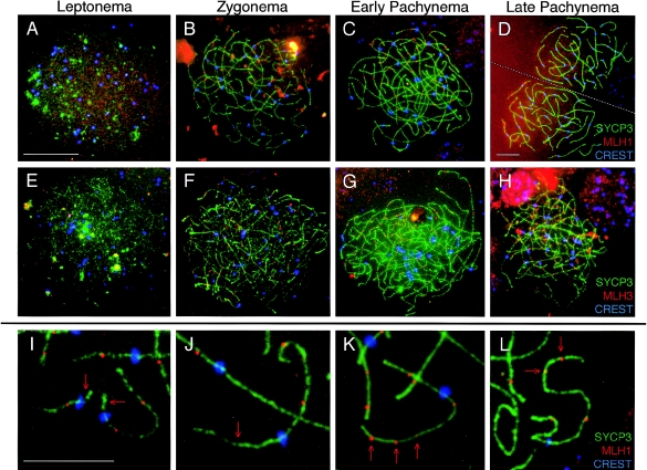
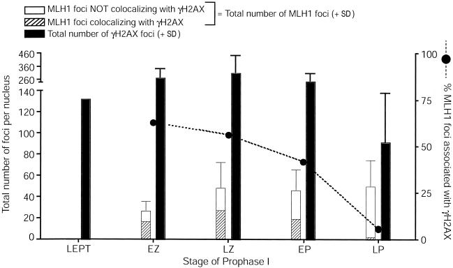
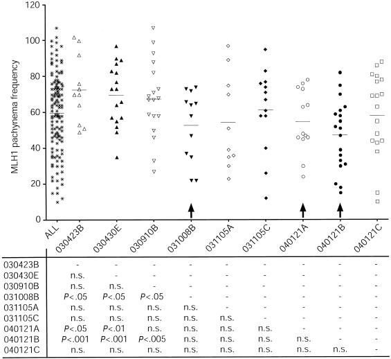
In humans, ~50% of conceptuses are chromosomally aneuploid as a consequence of errors in meiosis, and most of these aneuploid conceptuses result in spontaneous miscarriage. Of these aneuploidy events, 70% originate during maternal meiosis, with the majority proposed to arise as a direct result of defective crossing over during meiotic recombination in prophase I. By contrast, <1%-2% of mouse germ cells exhibit prophase I-related nondisjunction events. This disparity among mammalian species is surprising, given the conservation of genes and events that regulate meiotic progression. To understand the mechanisms that might be responsible for the high error rates seen in human females, we sought to further elucidate the regulation of meiotic prophase I at the molecular cytogenetic level. Given that these events occur during embryonic development in females, samples were obtained during a defined period of gestation (17-24 weeks). Here, we demonstrate that human oocytes enter meiotic prophase I and progress through early recombination events in a similar temporal framework to mice. However, at pachynema, when chromosomes are fully paired, we find significant heterogeneity in the localization of the MutL homologs, MLH1 and MLH3, among human oocyte populations. MLH1 and MLH3 have been shown to mark late-meiotic nodules that correlate well with--and are thought to give rise to--the sites of reciprocal recombination between homologous chromosomes, which suggests a possible 10-fold variation in the processing of nascent recombination events. If such variability persists through development and into adulthood, these data would suggest that as many as 30% of human oocytes are predisposed to aneuploidy as a result of prophase I defects in MutL homolog-related events.


Figures

Figure  1
Figure 1
Localization of γH2AX during prophase I in human fetal oocytes. A–D, γH2AX localizes to human female meiotic chromosomes during each substage of prophase I. Human oocyte chromosome “spreads” were subjected to immunofluorescent localization of SYCP3, a component of the SC (green, FITC), with γH2AX (red, TRITC) and CREST (blue, Cy5), at leptonema (A), zygonema (B), early pachynema (C), and late pachynema (D). In addition, γH2AX (red, TRITC) was colocalized with sites containing either RAD51 (E and F) or MLH1 (G and H). In panels E, F, G, and H, γH2AX (red, TRITC) is colocalized with RAD51 (green, FITC) and CREST (blue, Cy5) during leptonema (E) and zygonema (F). In panels G and H, γH2AX (red, TRITC) is colocalized with MLH1 (green, FITC) and CREST (blue, Cy5) during late zygonema (G) and pachynema (H). Scale bar = 10 μm.
Figure  2
Figure 2
Localization of DNA repair proteins during prophase I in human fetal oocytes. Graphs show quantitation (number of foci per nucleus + SD) for each substage of prophase I for RAD51 (A), RPA (B), MSH4 (C), MSH5 (D), MLH1 (E), and MLH3 (F). Examples of these localization patterns are provided in figures 3–5. LEPT = leptonema; EZ = early zygonema; LZ = late zygonema; EP = early pachynema; MP = mid-pachynema; LP = late pachynema. Data were analyzed by ANOVA after log transformation to ensure equal variances (P values are given within each bar graph; in all cases, P<.0001). Pairwise comparisons of substages were performed using the Tukey posttest and are displayed in table format below each graph. One asterisk (*) indicates P<.05, two asterisks (**) indicate P<.01, and three asterisks (***) indicate P<.001; n.s. = not significant.
Figure  3
Figure 3
Localization of RAD51 and RPA during prophase I in human fetal oocytes. Human oocyte chromosome “spreads” were subjected to immunofluorescent localization of RAD51 and RPA to identify their pattern of distribution during each substage of prophase I. A–D, For each stage, RAD51 (red, TRITC) is shown with SYCP3 (green, FITC) and CREST (blue, Cy5). A, Leptonema; B, zygonema; C, early pachynema; and D, late pachynema. E–H, RPA (red, TRITC) is shown with SYCP3 (green, FITC) and CREST (blue, Cy5) during leptonema (E), zygonema (F), early pachynema (G), and late pachynema (H). Scale bar = 10 μm.
Figure  4
Figure 4
Localization of MSH4 and MSH5 during prophase I in human fetal oocytes. The MutS homologs, MSH4 and MSH5, are present along human female meiotic chromosomes during each substage of prophase I. A–D, During each stage, MSH4 (red, TRITC) is shown with SYCP3 (green, FITC) and CREST (blue, Cy5). A, Leptonema; B, zygonema; C, early pachynema; and D, late pachynema. MSH5 (red, TRITC) is shown with SYCP3 (green, FITC) and CREST (blue, Cy5) during leptonema (E), zygonema (F), early pachynema (G), and late pachynema (H). Scale bar = 10 μm.
Figure  5
Figure 5
Localization of MLH1 and MLH3 during prophase I in human fetal oocytes. The MutL homologs, MLH1 and MLH3, localize to human female meiotic chromosomes during each substage of prophase I. A–D, MLH1 (red, TRITC) is shown with SYCP3 (green, FITC) and CREST (blue, Cy5) during leptonema (A), zygonema (B), early pachynema (C), and late pachynema (D). E–H, During each stage, MLH3 (red, TRITC) is shown with SYCP3 (green, FITC) and CREST (blue, Cy5). E, Leptonema; F, zygonema; G, early pachynema; and H, late pachynema. I–L, High magnification images of pachytene chromosomes highlights extreme variability in MLH1 localization, with some chromosome arms of pachytene oocytes often having too few MLH1 foci (arrows in I and J) or too many MLH1 foci (arrows in K and L). Green, SYCP3; red, MLH1; blue, CREST. Scale bar for panels A–C and E–H = 10 μm, scale bar for panel D = 10 μm, and scale bar for panels I–K = 5 μm. Note: at leptonema, γH2AX localization is almost continuous along the developing SC, so counts at this stage do not necessarily represent single foci.
Figure  6
Figure 6
MLH1 interaction with γH2AX-positive sites throughout prophase I in human fetal oocytes. Colocalization of MLH1 with sites that contain γH2AX. For each stage, total MLH1 foci are shown in the left bar, which is subdivided into those MLH1 foci that are associated with γH2AX (hatched portion of bar) and those that are independent of γH2AX (unhatched portion of bar). The error bar reflects the SD for total MLH1 focus numbers. The blackened circles and dashed line represent the total percentage of MLH1 foci that are associated with γH2AX. The total number of γH2AX foci (+ SD) is represented by the right blackened bar for each stage.
Figure  7
Figure 7
MLH1 localization during pachynema in oocyte pools from individual fetal ovary samples. Each column represents oocytes from an individual fetal ovary pair (gestational age 17–24 wk). The “ALL” column represents MLH1 counts in the entire pool of ovaries. Results from unpaired t tests are given below each column. Certain oocyte pools have significantly lower MLH1 focus frequencies (arrows); n.s. = not significant.

Similar articles

Cited by

References

Electronic-Database Information

    1. Online Mendelian Inheritance in Man (OMIM), http://www.ncbi.nlm.nih.gov/Omim/ (for Sycp3, RAD51, RPA, MSH4, MSH5, MLH1, MLH3, H2AX, and SPO11 clone)

References

    1. Abdullah MF, Hoffmann ER, Cotton VE, Borts RH (2004) A role for the MutL homologue MLH2 in controlling heteroduplex formation and in regulating between two different crossover pathways in budding yeast. Cytogenet Genome Res 107:180–19010.1159/000080596 - DOI - PubMed
    1. Allers T, Lichten M (2001) Intermediates of yeast meiotic recombination contain heteroduplex DNA. Mol Cell 8:225–23110.1016/S1097-2765(01)00280-5 - DOI - PubMed
    1. Ashley T, Plug AW, Xu J, Solari AJ, Reddy G, Golub EI, Ward DC (1995) Dynamic changes in Rad51 distribution on chromatin during meiosis in male and female vertebrates. Chromosoma 104:19–2810.1007/s004120050088 - DOI - PubMed
    1. Baker SM, Plug AW, Prolla TA, Bronner CE, Harris AC, Yao X, Christie DM, Monell C, Arnheim N, Bradley A, Ashley T, Liskay RM (1996) Involvement of mouse Mlh1 in DNA mismatch repair and meiotic crossing over. Nat Genet 13:336–34210.1038/ng0796-336 - DOI - PubMed
    1. Bannister LA, Schimenti JC (2004) Homologous recombinational repair proteins in mouse meiosis. Cytogenet Genome Res 107:191–20010.1159/000080597 - DOI - PubMed

Publication types

LinkOut - more resources