. 2005 Apr 15;387(Pt 2):497-506.
doi: 10.1042/BJ20041324.

Cleavage at the stem region releases an active ectodomain of the membrane type 1 matrix metalloproteinase

Affiliations

Cleavage at the stem region releases an active ectodomain of the membrane type 1 matrix metalloproteinase

Marta Toth et al. Biochem J. .

Abstract

MT1-MMP (membrane type 1 matrix metalloproteinase) is a membrane-anchored MMP that can be shed to the extracellular milieu. In the present study we report the primary structure and activity of the major soluble form of MT1-MMP. MS analysis of the purified 50-kDa soluble MT1-MMP form shows that the enzyme extends from Tyr112 to Val524, indicating that formation of this species requires a proteolytic cleavage within the stem region. In agreement, deletion of the entire stem region of MT1-MMP inhibited shedding of the 50-kDa species. A recombinant 50-kDa species (Tyr112-Val524) expressed in cells exhibited enzymatic activity against pro-MMP-2 and galectin-3, and thus this species is a competent protease. The recombinant 50-kDa soluble form also decreased the level of surface-associated TIMP-2 (tissue inhibitor of metalloproteinase 2) when administered to cells expressing wild-type membrane-anchored MT1-MMP, suggesting that ectodomain shedding of MT1-MMP can alter the MMP/TIMP balance on the cell surface. A approximately 53-kDa species of MT1-MMP was also isolated from a non-detergent extract of human breast carcinoma tissue and was found to lack the cytosolic tail, as determined with specific MT1-MMP domain antibodies. Together, these data show that MT1-MMP ectodomain shedding is a physiological process that may broaden MT1-MMP activity to the pericellular space.


Figures

Figure 1
Figure 1. Characterization of soluble MT1-MMP forms
(A) Domain structure of active MT1-MMP with the domain specific antibodies used in the present study. (B) Media from BS-C-1 cells expressing MT1-MMP in the absence (lane 1) or in the presence of 2 μM TAPI-1 (lanes 2–4) were concentrated and subjected to SDS/PAGE (15% reducing gel) followed by immunoblot analysis using the mAbLEM-2/15 to the catalytic domain (lanes 1 and 2), pAbCT to cytoplasmic tail (lane 3) and pAb437 to the haemopexinlike domain (lane 4) of MT1-MMP. (C) Media of MT1-MMP expressing BS-C-1 cells obtained in the presence of 2 μM TAPI-1 were collected (lane 5) and subjected to ultracentrifugation (supernatant, lane 6; pellet, lane 7). The samples were subjected to SDS/PAGE (15% reducing gel) followed by immunoblot analysis using the mAbLEM-2/15 to the catalytic domain (lanes 5 and 6) and pAb437 to the haemopexin-like domain (lane 7). (D) Summary of the species of soluble forms of MT1-MMP detected in the media of BS-C-1 cells expressing MT1-MMP and their recognition by various anti-MT1-MMP antibodies. Cat.dom., catalytic domain; CT, C-terminus; HLD, haemopexin-like domain; TM, transmembrane.
Figure 2
Figure 2. MT1-MMP forms shed by surface biotinylated HT1080 cells
(A) Serum-free conditioned media from PMA (‘TPA’)- (lane 1) or Con A-treated (lane 2) HT1080 cells were incubated with the MMP inhibitor-tethered matrix and the bound proteins were detected by immunoblot (IB) analysis using mAbLEM-2/15. (B) Untreated (lanes 1, 3, 5 and 7) or Con A-treated (lanes 2, 4, 6 and 8) HT1080 cells were surface biotinylated (lanes 3, 4, 7 and 8) or non-biotinylated (lanes 1, 2, 5 and 6). The serum-free conditioned media were collected, incubated with the inhibitor-tethered matrix and the bound fractions were subjected to SDS/PAGE (12% reducing gel) and transferred on to a nitrocellulose membrane. The same membrane was developed with streptavidin/HRP (lanes 1–4) and with mAbLEM-2/15 (lanes 5–8). (C) Serum-free media of MT1-MMP expressing BS-C-1 cells were incubated with the inhibitor-tethered matrix in the absence or presence of recombinant TIMP-2 or TIMP-1. The 50 kDa form of MT1-MMP in the unbound and bound fractions were detected by immunoblot analysis using the mAbLEM-2/15.
Figure 3
Figure 3. Isolation and structure of the 50-kDa soluble form of MT1-MMP
(A) Concentrated medium of MT1-MMP expressing BS-C-1 cells (lane 1 and 4) was incubated with the inhibitor-tethered matrix and the unbound (lanes 2 and 5) and bound fractions (lanes 3 and 6) were subjected to SDS/PAGE (12% reducing gel) followed by immunoblot (IB) analysis using mAbLEM-2/15 to the catalytic domain (lanes 1–3) or pAb437 to the haemopexin-like domain (lanes 4–6). (B) Structure of the 50-kDa soluble species of MT1-MMP as determined by MS/MS sequencing. The arrow indicates the putative site of cleavage at the stem region.
Figure 4
Figure 4. Mutations and truncations at the stem region of MT1-MMP: effect on shedding
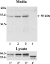
Concentrated media and cell lysates of CV-1 cells infected/transfected to express wild-type (lanes 1 and 1′), Ile525→Phe (lanes 2 and 2′), Val524→Glu/Ile525→Glu (lanes 3 and 3′) or Δstem (lanes 4 and 4′) MT1-MMP were subjected to immunoblot analysis using mAbLEM-2/15 to the catalytic domain of MT1-MMP.
Figure 5
Figure 5. Expression and activity of recombinant soluble 50-kDa MT1-MMP
(A) CV-1 cells were infected/transfected with (lanes 2 and 4) or without (lanes 1 and 3) a plasmid encoding the 50-kDa MT1-MMP as described in the Experimental section. The media and lysates were analysed by immunoblot using mAbLEM-2/15 to the catalytic domain. (B and C) CV-1 cells were infected/transfected to express wild-type MT1-MMP (lane 2) or the recombinant 50-kDa (Tyr112–Val524) soluble form (lane 3). Post-transfection (4 h), the media were replaced with serum-free media containing 10 nM pro-MMP-2 (B) or 20 nM galectin-3 (C) and incubated for an additional 18 h. An aliquot of the media was analysed by gelatin zymography (B) or by immunoblot analysis using an anti-galectin-3 antibody (C). The arrow in (B) indicates the intermediate form of MMP-2 and the asterisk in (C) indicates the cleaved product of galectin-3. Lanes 1 in (B) and (C) show the control pro-MMP-2 and control galectin-3 respectively.
Figure 6
Figure 6. Cellular distribution of TIMP-2 in the presence of membrane-anchored and soluble MT1-MMP
CV-1 cells were infected/transfected to express or not wild-type (wt) MT1-MMP. Post-transfection (4 h), the cells received 3.4 nM TIMP-2 alone or with ∼50 ng of recombinant 50-kDa MT1-MMP in serum-free media for 16 h. Then, the cell lysate and media were harvested and analysed for TIMP-2 levels by ELISA.
Figure 7
Figure 7. MMP inhibitor-tethered affinity isolation of MT1-MMP in extracts of benign and tumour breast tissues
Tumour and benign breast tissues isolated from six different human invasive breast carcinomas were homogenized, as described in the Experimental section, and incubated with the MMP inhibitor-tethered matrix. The bound protein fraction from tumour (lane 1) and benign (lane 2) tissue were eluted from the immobilized inhibitor, and subjected to immunoblot (IB) analysis of the same blot with either the mAbLEM-2/15 to the catalytic domain (A) or the pAbCT to the cytosolic tail (B) of MT1-MMP. Lane 3 shows the membrane-anchored (57 kDa) and soluble (50 kDa) MT1-MMP found in BS-C-1 cells as a control. Note that the ∼53-kDa form in the tumour tissue shown in (A) (lane 1) is only recognized by the antibody to the catalytic domain.

Similar articles

Cited by

References

    1. Hotary K. B., Allen E. D., Brooks P. C., Datta N. S., Long M. W., Weiss S. J. Membrane type I matrix metalloproteinase usurps tumor growth control imposed by the three-dimensional extracellular matrix. Cell. 2003;114:33–45. - PubMed
    1. Hotary K., Allen E., Punturieri A., Yana I., Weiss S. J. Regulation of cell invasion and morphogenesis in a three-dimensional type I collagen matrix by membrane-type matrix metalloproteinases 1, 2, and 3. J. Cell Biol. 2000;149:1309–1323. - PMC - PubMed
    1. Ohuchi E., Imai K., Fujii Y., Sato H., Seiki M., Okada Y. Membrane type 1 matrix metalloproteinase digests interstitial collagens and other extracellular matrix macromolecules. J. Biol. Chem. 1997;272:2446–2451. - PubMed
    1. Strongin A. Y., Collier I., Bannikov G., Marmer B. L., Grant G. A., Goldberg G. I. Mechanism of cell surface activation of 72-kDa type IV collagenase. Isolation of the activated form of the membrane metalloprotease. J. Biol. Chem. 1995;270:5331–5338. - PubMed
    1. Knauper V., Will H., Lopez-Otin C., Smith B., Atkinson S. J., Stanton H., Hembry R. M., Murphy G. Cellular mechanisms for human procollagenase-3 (MMP-13) activation. Evidence that MT1-MMP (MMP-14) and gelatinase a (MMP-2) are able to generate active enzyme. J. Biol. Chem. 1996;271:17124–17131. - PubMed

Publication types

Substances