. 2005 Feb 4;280(5):3847-61.
doi: 10.1074/jbc.M412299200. Epub 2004 Nov 22.

Molecular characterization, expression, and in vivo analysis of LmexCht1: the chitinase of the human pathogen, Leishmania mexicana

Affiliations

Molecular characterization, expression, and in vivo analysis of LmexCht1: the chitinase of the human pathogen, Leishmania mexicana

Manju B Joshi et al. J Biol Chem. .

Abstract

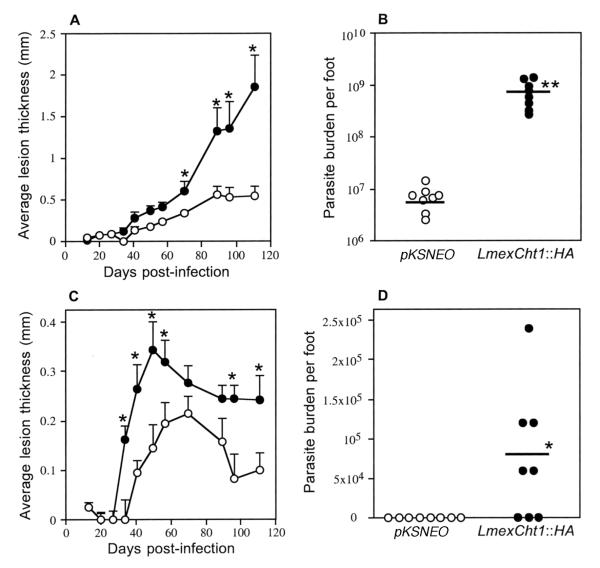
Chitinases have been implicated to be of importance in the life cycle development and transmission of a variety of parasitic organisms. Using a molecular approach, we identified and characterized the structure of a single copy LmexCht1-chitinase gene from the primitive trypanosomatid pathogen of humans, Leishmania mexicana. The LmexCht1 encodes an approximately 50 kDa protein, with well conserved substrate binding and catalytic domains characteristic of members of the chitinase-18 protein family. Further, we showed that LmexCht1 mRNA is constitutively expressed by both the insect vector (i.e. promastigote) and mammalian (i.e. amastigote) life cycle developmental forms of this protozoan parasite. Interestingly, however, amastigotes were found to secrete/release approximately >2-4-fold higher levels of chitinase activity during their growth in vitro than promastigotes. Moreover, a homologous episomal expression system was devised and used to express an epitope-tagged LmexCht1 chimeric construct in these parasites. Expression of the LmexCht1 chimera was verified in these transfectants by reverse transcription-PCR, Western blots, and indirect immunofluorescence analyses. Further, results of coupled immunoprecipitation/enzyme activity experiments demonstrated that the LmexCht1 chimeric protein was secreted/released by these transfected L. mexicana parasites and that it possessed functional chitinase enzyme activity. Such transfectants were also evaluated for their infectivity both in human macrophages in vitro and in two different strains of mice. Results of those experiments demonstrated that the LmexCht1 transfectants survived significantly better in human macrophages and also produced significantly larger lesions in mice than control parasites. Taken together, our results indicate that the LmexCht1-chimera afforded a definitive survival advantage to the parasite within these mammalian hosts. Thus, the LmexCht1 could potentially represent a new virulence determinant in the mammalian phase of this important human pathogen.


Figures

FIG. 1
FIG. 1. The sequence and structure of the L. mexicana chitinase
A. The deduced amino acid sequence of the L. mexicana chitinase 1 (LmexCht1) gene. The underlined sequence delineates a putative 28 aa signal peptide (Met1 – Ser28). The light gray boxes (marked: I [Leu123-Ala139] and III [Leu249-Gly264]) indicate the two putative substrate binding sites and the open box (marked: II [Arg150- Thr179]) indicate the putative catalytic/active site of this enzyme. The bold-italicized residues within the open box (Leu167- Glu175) represent the signature sequence of the Chitinase-18 Protein Family. The two potential N-linked glycosylation sites (Asn48 and Asn384) are marked with an asterisk and the single potential O-linked glycosylation site (Thr52) is indicated by (τ). The dashed-underlined aa sequences (designated as Pep1-Pep4) represent regions with high levels of conservation to antigenic peptide epitopes in the L. donovani Cht1 deduced protein. B. Comparison of the conserved functional domains of the L. mexicana Cht1 (L. mex.) and L. donovani Cht1 (L.d.) chitinases. The conserved substrate binding sites (i.e. Regions I and III) and the conserved catalytic/active site (i.e. Region II) are indicated. The divergent aa residues between these two proteins are shown in bold-face.
FIG. 2
FIG. 2. Southern Blot analyses of the L. mexicana chitinase 1 gene locus
A. Southern hybridization of L. mexicana gDNA with the LmexCht1-DIG1151 probe. gDNA (5 μg) was digested with various individual restriction endonucleases ( EcoRI, Hind III, Apa I, Not I, BamH I, Sph I and Mlu I, as indicated), separated in 1% agarose gel, transferred to a nylon membrane and hybridized with the digoxigenin-labeled LmexCht1-DIG1151probe (i.e. corresponding to nt 1 - nt 1151 of the LmexCht1-ORF). DNA standards (in bp) are shown on the left. B. Southern hybridization of the Lmex-Cos1 cosmid DNA with the LmexCht1-DIG1151 probe. Cosmid DNA (1 μg) was digested with the same restriction endonucleases as shown in Panel A and subjected to Southern hybridization with the LmexCht1-DIG1151 probe. DNA standards (in bp) are shown on the left as in Panel A.
FIG. 3
FIG. 3. Northern analyses and mapping of the spliced-leader acceptor site
A. Northern blot analysis of LmexCht1 mRNA transcripts present in various L. mexicana parasite developmental forms. Top Panel. Total RNA (5 μg) isolated from L. mexicana mouse lesion amastigotes (i.e. in vivo-derived), axenic amastigotes (i.e. in vitro-grown), and log (procyclic)- and stationary (metacyclic)-phase promastigotes was separated in an agarose gel, transferred onto a nylon membrane and hybridized with the LmexCht1-DIG 270 probe (i.e. corresponding to nt 502– nt 771 of the LmexCht1-ORF). Arrow indicates the position of the ~3 kb LmexCht1 mRNA transcript in these samples. Bottom Panel. Ethidium bromide strained agarose gel (used in panel A) showing the ribosomal RNA in each of the total RNA samples used above. B. Mapping of the LmexCht1 5′-spliced-leader acceptor site. Nucleotide sequence of the RT-PCR product obtained with LmexCht1 mRNA amplified with an L. mexicana-spliced leader (forward: SpliceFwd) oligonucleotide primer and an internal LmexCht1 (reverse: ORF-RT/Rev) primer. The 5′-untranslated region of the LmexCht1 gene is shown in lower case letters. The LmexCht1 open reading frame is shown in Upper case letters, a portion of the conserved spliced leader sequence (i.e. forward primer) is shown in underlined Caps and the arrow (↓) marks the position of the spliced-leader acceptor site. The ATG-start codon of the LmexCht1 open reading frame is double-underlined.
FIG. 4
FIG. 4. Episomal expression of the LmexCht1∷HA chimera in L. mexicana promastigotes
A. Map of the LmexCht1∷HA chimeric construct. A schematic representation showing the complete open reading frame (i.e. nt 1 – nt 1371, minus the terminal TAG stop codon) of the LmexCht1 gene fused at its 3′-end with a 27-nt sequence encoding the hemagglutinin (HA) epitope (light gray box). The black box at the 5′-end represents nt-1 to nt-84 encoding the putative 28 aa signal peptide (SP) of the LmexCht1 protein. The thick black line represents the pKSNEO plasmid (leishmanial) expression vector, and the Spe I restriction endonuclease sites used for cloning are shown. B. Growth kinetics of L. mexicana transfectants in vitro. The growth kinetics of promastigotes transfected with either LmexCht1∷HA (●) or pKSNEO control (○) constructs were monitored for their growth in vitro over a period of five days. Quadruplicate cultures were initiated at ~1-2 × 10⁶ cells ml−1 and aliquots taken at various time points for cell counting. Values shown represent the mean of three separate determinations for each culture.
FIG. 5
FIG. 5. RT-PCR analysis of episomally expressed NEO and LmexCht1∷HA mRNAs
A. Schematic representations of the pKSNEO control plasmid and the LmexCht1∷HA construct. 1. The pKSNEO plasmid: the dark gray box represents the nt sequence encoding neomycin phosphotransferase (NEO), the black line denotes the pKSNEO plasmid vector backbone and Spe I indicates the restriction site used for insert cloning. 2. The LmexCht1∷HA construct: the black and white boxes represent nt-1 to nt-84 encoding the putative signal peptide and nt 85 - nt 1371 of the LmexCht1 ORF, respectively. The light gray box represents the nt sequence encoding the hemagglutinin (HA) epitope fused in-frame with the LmexCht1 ORF followed by a terminal TGA stop codon. The Spe I restriction sites used for cloning this insert are indicated. The dark gray box and black lines represent NEO and the vector backbone as in 1, above. Arrowheads show the positions of the primers used for amplification, the dashed lines and numbers denote the predicted size (in bp) of the PCR products. B. A portion of an ethidium bromide stained agarose gel showing the amplification products obtained in RT-PCR. RNA isolated from L. mexicana promastigotes and (axenic) amastigotes transfected with either the pKSNEO control plasmid (lanes 2 and 4) or the LmexCht1∷HA (Cht1∷HA) construct (lanes 1 and 3) was reverse transcribed using oligo(dT). Aliquots of the resulting cDNAs were subjected to PCR amplification using the primer pairs shown above. Top panel: shows the resulting 324 bp product (← ΔNEO) amplified from the neomycin phosphotransferase gene present in the pKSNEO plasmid backbone of all transfectants. Bottom panel: the 979 bp product (← ΔCht1HA) amplified from LmexCht1∷HA transfectants.
FIG. 6
FIG. 6. Episomal expression of the LmexCht1∷HA chimeric protein in L. mexicana transfectants
A. Western blot of whole cell lysates of L. mexicana promastigotes and (axenic) amastigotes transfected with either the LmexCht1∷HA construct (Cht1∷HA) (lanes 1 and 3) or the pKSNEO control plasmid (lanes 2 and 4), probed with a mouse anti-HA monoclonal antibody. Molecular mass standards (in kDa) are shown on the left. Arrow denotes the ~50 kDa LmexCht1∷HA chimeric protein. B. Indirect immunofluorescence images of LmexCht1∷HA transfected promastigotes (panel 1) and axenic amastigotes (panel 2) probed with a primary mouse anti-HA monoclonal antibody and a FITC-labeled goat anti-mouse secondary antibody. F-denotes the anterior, flagellar end of promastigotes. Bar represents 2μm. C. Immunoprecipitates from culture supernatants of L. mexicana promastigotes and (axenic) amastigotes transfected with either the LmexCht1∷HA construct (Cht1∷HA) (lanes 1 and 3) or the pKSNEO control plasmid (lanes 2 and 4) were obtained using a rabbit anti-LdCht1-peptide antibody. Such immune complexes were separated in SDS-PAGE, transblotted onto PVDF membranes and probed with a mouse anti-HA monoclonal antibody. Molecular mass standards (in kDa) are shown on the left. Arrow denotes the ~50 kDa LmexCht1∷HA chimeric protein.
FIG. 7
FIG. 7. Survival of L. mexicana transfectants in human macrophages in vitro
Human monocyte-derived macrophages were incubated with stationary phase L. mexicana promastigotes, transfected with either the LmexCht1∷HA chimeric construct or the pKSNEO control plasmid at a parasite to host cell ratio of 10:1. After 5h of incubation, extracellular (free) parasites were removed by aspiration. Following this, one set of cultures was immediately fixed for light microscopy and a second set was incubated for an additional 72 h prior to fixation. Subsequently, both sets of cultures were stained and examined by light microscopy. A. Percentage of macrophages infected by L. mexicana parasites transfected with either the LmexCht1∷HA chimeric construct (∎) or the pKSNEO control plasmid (□) after 72 h post-infection, respectively. B. Total number of intracellular parasites (i.e. amastigotes) per 100 macrophages after 72h post-infection with either LmexCht1∷HA (∎) or the pKSNEO (□) control transfectants. The data shown are from a single experiment but are typical of those obtained from two separate and independent macrophage infection experiments. The values given represent the mean ± SD obtained from triplicate samples analyzed at each time point. Statistically significant differences between experimental and control groups are as indicated (* p < 0.01 and ** p < 0.001).
FIG. 8
FIG. 8. Infectivity of L. mexicana transfectants in mice
Highly susceptible BALB/c (Panel A) and more resistant CBA/Ca (Panel C) mice (8 animals per group) were injected with 500 metacyclic promastigotes of either LmexCht1∷HA (●) or the pKSNEO control (○) transfectants into the dorsal surface of the right hind foot. Lesion development was monitored weekly by measuring the difference in thickness between the infected and the uninfected foot. The data shown are from a single experiment but are typical of those obtained from two separate and independent mouse infection experiments. The values given represent the mean ± SE obtained from each group of infected mice. Asterisk (*) above error bars represent statistically significant differences (p < 0.05) between experimental and control groups. After 15 weeks post-infection, the parasite burden in each individual BALB/c (Panel B) and CBA/Ca (Panel D) mouse foot-lesion was determined following homogenization and direct microscopic counting of released parasites (amastigotes). Horizontal bars represent the mean parasite burden determined for each group (n= 8) of infected animals. Statistically significant differences between experimental and control groups are indicated (* p < 0.05 and ** p < 0.0005).

Similar articles

Cited by

References

    1. UNDP/WorldBank/WorldHealthOrganization . Tropical Disease Resaerch: Progress 1997-1998: Fourteenth Programme Report of UNDP/World Bank/WHO Special Programme for Research and Training in Tropical Diseases. World Health Organization; Geneva, Switzerland: 1999.
    1. Handman E. Clin Microbiol Rev. 2001;14:229–243. - PMC - PubMed
    1. Sacks D, Kamhawi S. Annu Rev Microbiol. 2001;55:453–483. - PubMed
    1. Schlein Y, Jacobson RL, Shlomai J. Proc R Soc Lond B Biol Sci. 1991;245:121–126. - PubMed
    1. Schlein Y, Jacobson RL, Messer G. Proc Natl Acad Sci U S A. 1992;89:9944–9948. - PMC - PubMed

Publication types

MeSH terms

Associated data

LinkOut - more resources