. 2004 Dec;2(12):e427.
doi: 10.1371/journal.pbio.0020427. Epub 2004 Nov 30.

A transcriptional profile of aging in the human kidney

Affiliations

A transcriptional profile of aging in the human kidney

Graham E J Rodwell et al. PLoS Biol. 2004 Dec.

Abstract

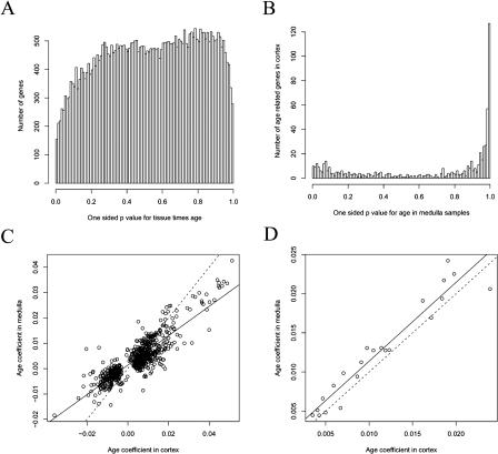
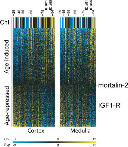
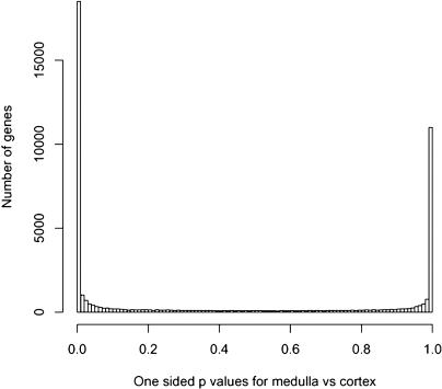
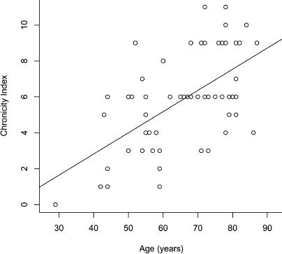
In this study, we found 985 genes that change expression in the cortex and the medulla of the kidney with age. Some of the genes whose transcripts increase in abundance with age are known to be specifically expressed in immune cells, suggesting that immune surveillance or inflammation increases with age. The age-regulated genes show a similar aging profile in the cortex and the medulla, suggesting a common underlying mechanism for aging. Expression profiles of these age-regulated genes mark not only age, but also the relative health and physiology of the kidney in older individuals. Finally, the set of aging-regulated kidney genes suggests specific mechanisms and pathways that may play a role in kidney degeneration with age.


Conflict of interest statement

The authors have declared that no conflicts of interest exist.

Figures

Figure 1
Figure 1. Age-Regulated Genes
(A) Shown are expression levels for gene CDO1. White and black circles represent expression from cortex and medulla, respectively. The y-axis indicates log2 (expression level), and the x-axis indicates age of patient (years). Dotted and solid lines indicate best fit slopes for the cortex and medulla values, respectively. (B) For every gene, we calculated a one-sided p˜ -value that its expression changes with age. Shown is a histogram representing all of the genes represented by the Affymetrix DNA chip. Genes that decrease with age have p˜ -values near zero, and genes that increase with age have p˜ -values near one. If there were no age-regulated genes (i.e., the true βkj = 0 for every gene j), then the histogram of p˜ -values would be flat (i.e., have a uniform distribution on the interval from zero to one). The x-axis shows the p˜ -value, and the y-axis shows the number of genes with that p˜ -value. There are 985 genes with a p-value less than 0.001.
Figure 2
Figure 2. Similar Age-Regulation in Cortex and Medulla
(A) For every gene, we calculated a p˜ -value that there is a Tissuei ×Agei effect, and plotted the results in a histogram. Genes that show different age regulation in the cortex or the medulla would be contained in peaks on the left and right parts of the histogram. The figure shows that the number of genes that have different expression levels in the cortex and medulla is about the same as or less than would be expected by chance. The x-axis shows one-sided p˜ -values for Tissuei ×Agei, and the y-axis shows the number of genes with that p˜ -value. There is a systematic under-representation of the edge regions compared to a random sample of uniform random variables because of correlations among the 44,928 p˜ -values computed from 133 samples. (B) To show whether aging in the cortex and the medulla is similar, we selected age-regulated genes in the cortex and calculated the one-tailed p˜ -value for age effects in the medulla. The histogram shows these selected p˜ -values. The spike at the right shows genes that increase with age in the medulla. Those genes also increased with age in the cortex. (C) Shown is a scatterplot of all 684 genes that are age-regulated in either the medulla or the cortex (p < 0.001). The y-axis is the slope for the medulla of the expression change with respect to age, and the x-axis is the slope for the cortex. The solid line is the least squares line, with a slope of 0.58. The dotted line has a slope of one and passes through the origin. (D) Same as (C) but for 22 genes that are age-regulated in both the cortex and the medulla (p < 0.001).
Figure 3
Figure 3. Expression of the 447 Genes as a Function of Age
Rows correspond to age-regulated genes, ordered from most highly induced to most highly repressed. Columns correspond to individual patients, ordered from youngest to oldest. The age of certain patients is shown for reference. Left panel refers to data from cortex samples, and right panel depicts data from medulla samples. The first row shows the chronicity index (ChI; morphological appearance and physiological state of the kidney),from blue (healthiest) to yellow (least healthy) as indicated in the scale bar. Key genes discussed in the text are marked. Scale shows log2 of the expression level (Exp). A navigable version of this figure can be found at http://cmgm.stanford.edu/~kimlab/aging_kidney/explorer.html.
Figure 4
Figure 4. Differential Expression in the Cortex and the Medulla
For each gene, we calculated a p˜ -value for expression differences in the cortex versus the medulla. Shown is a histogram of these p˜ -values. Genes enriched in the cortex are in a peak on the left, and genes enriched in the medulla are in a peak on the right. The x-axis indicates p˜ -value, and the y-axis indicates number of genes.
Figure 5
Figure 5. Developmental Profile of the Age-Regulated Genes
Shown are the log2 of the expression levels for 227 age-regulated genes in 26 human tissues, using data from Su et al. (2002). Rows correspond to genes, columns correspond to human tissues. a, kidney; b, cerebellum; c, whole brain; d, cerebral cortex; e, caudate nucleus; f, amygdala; g, thalamus; h, corpus callosum; i, spinal cord; j, whole blood; k, testis; l, pancreas; m, placenta; n, pituitary gland; o, thyroid gland; p, prostate; q, ovary; r, uterus; s, salivary gland; t, trachea; u, lung; v, thymus; w, spleen; x, adrenal gland; y, liver; z, heart. Scale shows log2 of the expression level. A navigable version of this figure can be found at http://cmgm.stanford.edu/~kimlab/aging_kidney/explorer.html.
Figure 6
Figure 6. Chronicity Index of Kidney Samples
Histology from patient 40 is shown on the left, demonstrating a normal glomerulus (G), tubules and interstitial space (T), and arteriole (A), respectively (chronicity score of zero). Histology from patient 62 is shown on the right, demonstrating glomerulosclerosis (g), tubular atrophy and interstitial fibrosis (t), and arterial intimal hyalinosis (a), respectively (chronicity score of ten). Hematoxylin and eosin staining of paraffin-embedded sections.
Figure 7
Figure 7. Chronicity Index Increases with Age
Shown is the chronicity index versus age for most of the kidney samples used in this study. The line shows the least squared fit through the data points.

References

    1. Droge W. Oxidative stress and aging. Adv Exp Med Biol. 2003;543:191–200. - PubMed
    1. Guarente L, Kenyon C. Genetic pathways that regulate ageing in model organisms. Nature. 2000;408:255–262. - PubMed
    1. Hasty P, Campisi J, Hoeijmakers J, van Steeg H, Vijg J. Aging and genome maintenance: Lessons from the mouse? Science. 2003;299:1355–1359. - PubMed
    1. Hekimi S, Guarente L. Genetics and the specificity of the aging process. Science. 2003;299:1351–1354. - PubMed
    1. Higgins JP, Shinghal R, Gill H, Reese JH, Terris M, et al. Gene expression patterns in renal cell carcinoma assessed by complementary DNA microarray. Am J Pathol. 2003;162:925–932. - PMC - PubMed

Publication types

MeSH terms