

Mucin glycosylation changes in cystic fibrosis lung disease are not manifest in submucosal gland secretions
- PMID: 15563276
- PMCID: PMC1135025
- DOI: 10.1042/BJ20041641
Mucin glycosylation changes in cystic fibrosis lung disease are not manifest in submucosal gland secretions
Abstract
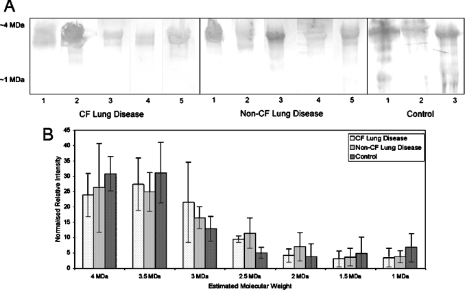
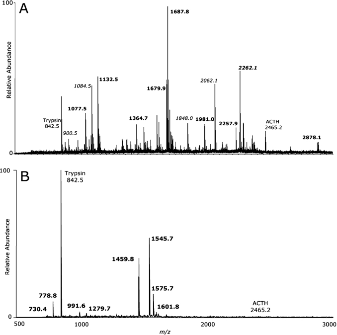
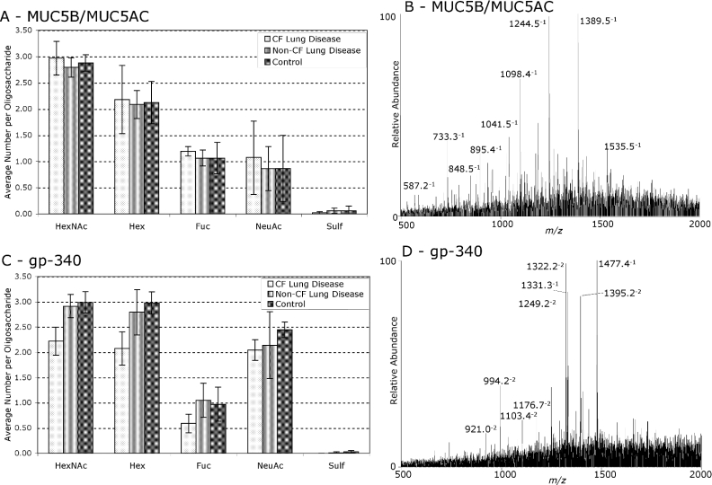
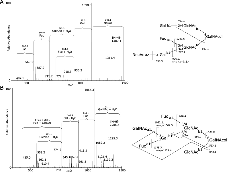
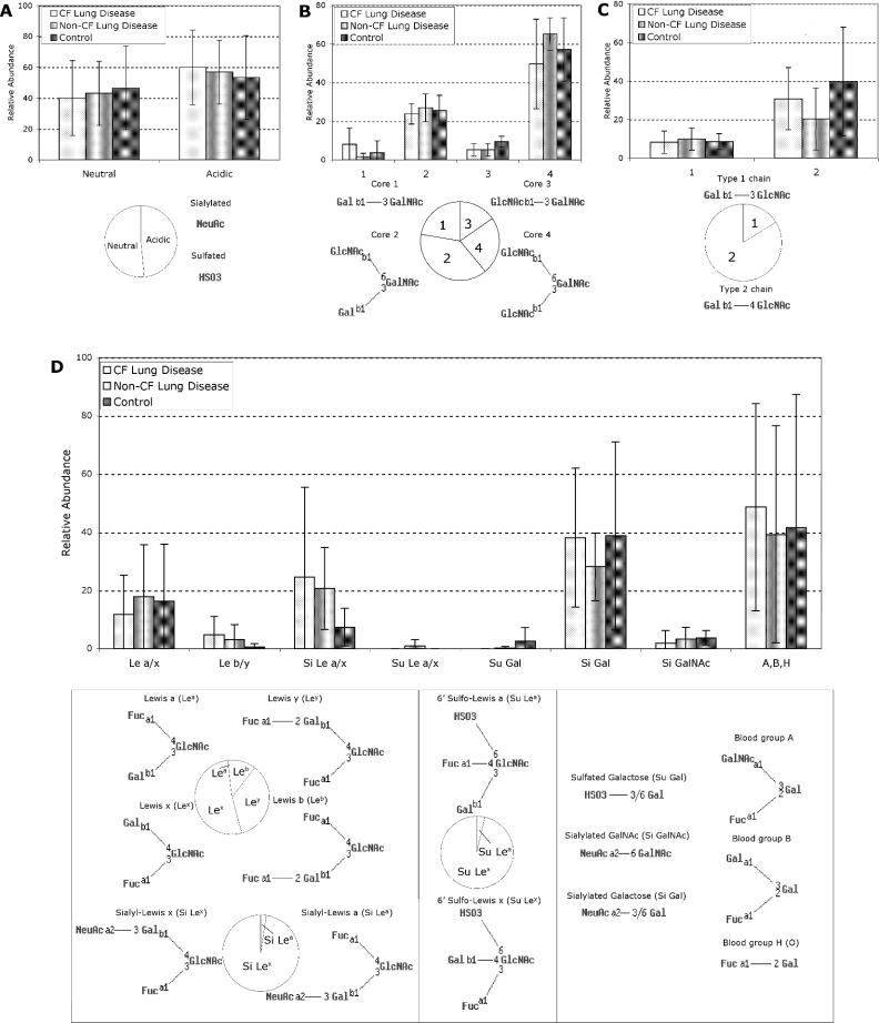
SMG (submucosal gland) secretions are a major component of the airway surface liquid, are associated with innate immunity in the lung, and have been reported to be altered in lung disease. Changes in lung mucosal glycosylation have been reported in CF (cystic fibrosis), which may be responsible for differential bacterial binding to glycosylated components in the lung mucosa and hence increased pre-disposition to pulmonary infection. Glycoproteomic analysis was performed on SMG secretions collected from explanted bronchial tissue of subjects with severe lung disease, with and without CF, and controls without lung disease. Mucins MUC5B and MUC5AC were shown to be the dominant high-molecular-mass glycoprotein components, with a minor non-mucin glycoprotein component, gp-340, also present. Oligosaccharides containing blood-group determinants corresponding to subjects' blood type were abundant on MUC5B/MUC5AC, as were Lewis-type epitopes and their sialylated analogues, which are ligands for pathogens and leucocytes. No significant differences were found in the glycosylation of MUC5B/MUC5AC or gp-340 between CF and non-CF subjects with severe lung disease, implying that CF does not influence SMG secretion mucin glycosylation in end-stage lung disease. There were also no significant differences found in the glycosylation of these components in severe lung disease compared with non-diseased lungs. This suggests that previously reported changes in the glycosylation of respiratory glycoconjugates in CF, and other pulmonary conditions, are not due to the glycosylation of components in SMG secretions, but may involve other secretions, responses or extracellular factors.
Figures
Similar articles
-
MUC5AC and MUC5B Mucins Are Decreased in Cystic Fibrosis Airway Secretions.Am J Respir Cell Mol Biol. 2004 Jul;31(1):86-91. doi: 10.1165/rcmb.2003-0345OC. Epub 2004 Feb 26. Am J Respir Cell Mol Biol. 2004. PMID: 14988081
-
Glycosylation of sputum mucins is altered in cystic fibrosis patients.Glycobiology. 2007 Jul;17(7):698-712. doi: 10.1093/glycob/cwm036. Epub 2007 Mar 28. Glycobiology. 2007. PMID: 17392389
-
MUC5AC and MUC5B mucins increase in cystic fibrosis airway secretions during pulmonary exacerbation.Am J Respir Crit Care Med. 2007 Apr 15;175(8):816-21. doi: 10.1164/rccm.200607-1011OC. Epub 2007 Jan 25. Am J Respir Crit Care Med. 2007. PMID: 17255563
-
Human airway mucin glycosylation: a combinatory of carbohydrate determinants which vary in cystic fibrosis.Glycoconj J. 2001 Sep;18(9):661-84. doi: 10.1023/a:1020867221861. Glycoconj J. 2001. PMID: 12386453 Review.
-
Mucin Secretion in Cystic Fibrosis: A Systematic Review.Dig Dis. 2021;39(4):375-381. doi: 10.1159/000512268. Epub 2020 Oct 13. Dig Dis. 2021. PMID: 33049746
Cited by
-
Mucins: the frontline defence of the lung.Biochem Soc Trans. 2018 Oct 19;46(5):1099-1106. doi: 10.1042/BST20170402. Epub 2018 Aug 28. Biochem Soc Trans. 2018. PMID: 30154090 Free PMC article. Review.
-
Cellular and molecular biology of airway mucins.Int Rev Cell Mol Biol. 2013;303:139-202. doi: 10.1016/B978-0-12-407697-6.00004-0. Int Rev Cell Mol Biol. 2013. PMID: 23445810 Free PMC article. Review.
-
Salivary mucin MUC7 oligosaccharides in patients with recurrent aphthous stomatitis.Clin Oral Investig. 2015 Nov;19(8):2147-52. doi: 10.1007/s00784-015-1495-3. Epub 2015 Jun 9. Clin Oral Investig. 2015. PMID: 26051835 Clinical Trial.
-
Effects of Muclin (Dmbt1) deficiency on the gastrointestinal system.Am J Physiol Gastrointest Liver Physiol. 2008 Mar;294(3):G717-27. doi: 10.1152/ajpgi.00525.2007. Epub 2008 Jan 17. Am J Physiol Gastrointest Liver Physiol. 2008. PMID: 18202109 Free PMC article.
-
COPD immunopathology.Semin Immunopathol. 2016 Jul;38(4):497-515. doi: 10.1007/s00281-016-0561-5. Epub 2016 May 13. Semin Immunopathol. 2016. PMID: 27178410 Free PMC article. Review.
References
-
- Sharma P., Dudus L., Nielsen P. A., Clausen H., Yankaskas J. R., Hollingsworth M. A., Engelhardt J. F. MUC5B and MUC7 are differentially expressed in mucous and serous cells of submucosal glands in human bronchial airways. Am. J. Respir. Cell Mol. Biol. 1998;19:30–37. - PubMed
-
- Finkbeiner W. E. Physiology and pathology of tracheobronchial glands. Respir. Physiol. 1999;118:77–83. - PubMed
-
- Jeffery P. K. Remodeling in asthma and chronic obstructive lung disease. Am. J. Respir. Crit. Care Med. 2001;164:S28–S38. - PubMed
Publication types
MeSH terms
Substances
LinkOut - more resources
Full Text Sources
Medical