

Vaccination with EphA2-derived T cell-epitopes promotes immunity against both EphA2-expressing and EphA2-negative tumors
- PMID: 15563374
- PMCID: PMC535538
- DOI: 10.1186/1479-5876-2-40
Vaccination with EphA2-derived T cell-epitopes promotes immunity against both EphA2-expressing and EphA2-negative tumors
Abstract
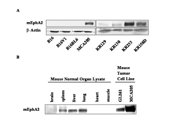
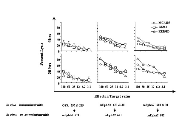
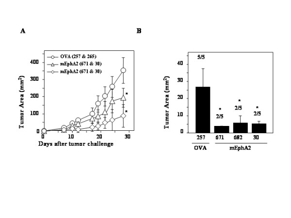
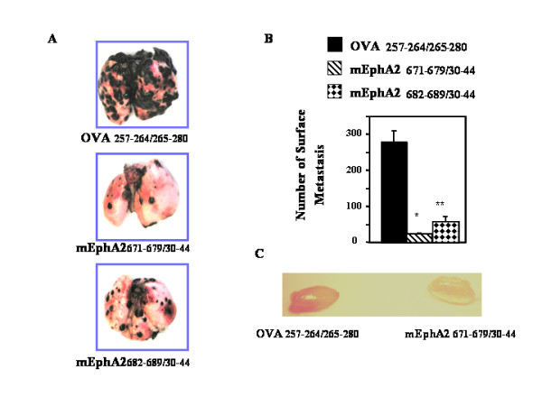
BACKGROUND: A novel tyrosine kinase receptor EphA2 is expressed at high levels in advanced and metastatic cancers. We examined whether vaccinations with synthetic mouse EphA2 (mEphA2)-derived peptides that serve as T cell epitopes could induce protective and therapeutic anti-tumor immunity. METHODS: C57BL/6 mice received subcutaneous (s.c.) vaccinations with bone marrow-derived dendritic cells (DCs) pulsed with synthetic peptides recognized by CD8+ (mEphA2671-679, mEphA2682-689) and CD4+ (mEphA230-44) T cells. Splenocytes (SPCs) were harvested from primed mice to assess the induction of cytotoxic T lymphocyte (CTL) responses against syngeneic glioma, sarcoma and melanoma cell lines. The ability of these vaccines to prevent or treat tumor (s.c. injected MCA205 sarcoma or B16 melanoma; i.v. injected B16-BL6) establishment/progression was then assessed. RESULTS: Immunization of C57BL/6 mice with mEphA2-derived peptides induced specific CTL responses in SPCs. Vaccination with mEPhA2 peptides, but not control ovalbumin (OVA) peptides, prevented the establishment or prevented the growth of EphA2+ or EphA2-negative syngeneic tumors in both s.c. and lung metastasis models. CONCLUSIONS: These data indicate that mEphA2 can serve as an attractive target against which to direct anti-tumor immunity. The ability of mEphA2 vaccines to impact EphA2-negative tumors such as the B16 melanoma may suggest that such beneficial immunity may be directed against alternative EphA2+ target cells, such as the tumor-associated vascular endothelial cells.
Figures
Similar articles
-
Immunotherapy of murine colon cancer using receptor tyrosine kinase EphA2-derived peptide-pulsed dendritic cell vaccines.Cancer. 2007 Oct 1;110(7):1469-77. doi: 10.1002/cncr.22958. Cancer. 2007. PMID: 17685394
-
Potent CD4+ T-cell epitope P30 enhances HER2/neu-engineered dendritic cell-induced immunity against Tg1-1 breast cancer in transgenic FVBneuN mice by enhanced CD4+ T-cell-stimulated CTL responses.Cancer Gene Ther. 2013 Oct;20(10):590-8. doi: 10.1038/cgt.2013.60. Epub 2013 Sep 20. Cancer Gene Ther. 2013. PMID: 24052129
-
Dendritic cells break tolerance and induce protective immunity against a melanocyte differentiation antigen in an autologous melanoma model.Cancer Res. 2000 Dec 15;60(24):6995-7001. Cancer Res. 2000. PMID: 11156402
-
[Anti-metastatic effect of vascular endothelial growth factor receptor 2 extracellular domain gene-modified dendritic cell vaccination in murine model with experimental pulmonary metastasis].Zhonghua Zhong Liu Za Zhi. 2006 Sep;28(9):646-9. Zhonghua Zhong Liu Za Zhi. 2006. PMID: 17274366 Chinese.
-
Natural CD8⁺25⁺ regulatory T cell-secreted exosomes capable of suppressing cytotoxic T lymphocyte-mediated immunity against B16 melanoma.Biochem Biophys Res Commun. 2013 Aug 16;438(1):152-5. doi: 10.1016/j.bbrc.2013.07.044. Epub 2013 Jul 20. Biochem Biophys Res Commun. 2013. PMID: 23876314
Cited by
-
Immunotherapeutic approaches for glioma.Crit Rev Immunol. 2009;29(1):1-42. doi: 10.1615/critrevimmunol.v29.i1.10. Crit Rev Immunol. 2009. PMID: 19348609 Free PMC article. Review.
-
Ephs in cancer progression: complexity and context-dependent nature in signaling, angiogenesis and immunity.Cell Commun Signal. 2024 May 29;22(1):299. doi: 10.1186/s12964-024-01580-3. Cell Commun Signal. 2024. PMID: 38811954 Free PMC article. Review.
-
Eph Receptor Tyrosine Kinases in Tumor Immunity.Cancer Res. 2016 Nov 15;76(22):6452-6457. doi: 10.1158/0008-5472.CAN-16-1521. Epub 2016 Nov 3. Cancer Res. 2016. PMID: 27811149 Free PMC article. Review.
-
Toll like receptor-3 ligand poly-ICLC promotes the efficacy of peripheral vaccinations with tumor antigen-derived peptide epitopes in murine CNS tumor models.J Transl Med. 2007 Feb 12;5:10. doi: 10.1186/1479-5876-5-10. J Transl Med. 2007. PMID: 17295916 Free PMC article.
-
Impact of combination immunochemotherapies on progression of 4NQO-induced murine oral squamous cell carcinoma.Cancer Immunol Immunother. 2019 Jul;68(7):1133-1141. doi: 10.1007/s00262-019-02348-2. Epub 2019 May 28. Cancer Immunol Immunother. 2019. PMID: 31139925 Free PMC article.
References
-
- Zelinski DP, Zantek ND, Stewart JC, Irizarry AR, Kinch MS. EphA2 overexpression causes tumorigenesis of mammary epithelial cells. Cancer Res. 2001;61:2301–2306. - PubMed
-
- Easty DJ, Guthrie BA, Maung K, Farr CJ, Lindberg RA, Toso RJ, Herlyn M, Bennett DC. Protein B61 as a new growth factor: expression of B61 and up-regulation of its receptor epithelial cell kinase during melanoma progression. Cancer Res. 1995;55:2528–2532. - PubMed
-
- Easty DJ, Hill SP, Hsu MY, Fallowfield ME, Florenes VA, Herlyn M, Bennett DC. Up-regulation of ephrin-A1 during melanoma progression. Int J Cancer. 1999;84:494–501. - PubMed
Grants and funding
LinkOut - more resources
Full Text Sources
Other Literature Sources
Molecular Biology Databases
Research Materials
Miscellaneous
