. 2005 Feb 25;280(8):6960-8.
doi: 10.1074/jbc.M412752200. Epub 2004 Nov 24.

The heteromeric organic solute transporter alpha-beta, Ostalpha-Ostbeta, is an ileal basolateral bile acid transporter

Affiliations

The heteromeric organic solute transporter alpha-beta, Ostalpha-Ostbeta, is an ileal basolateral bile acid transporter

Paul A Dawson et al. J Biol Chem. .

Abstract

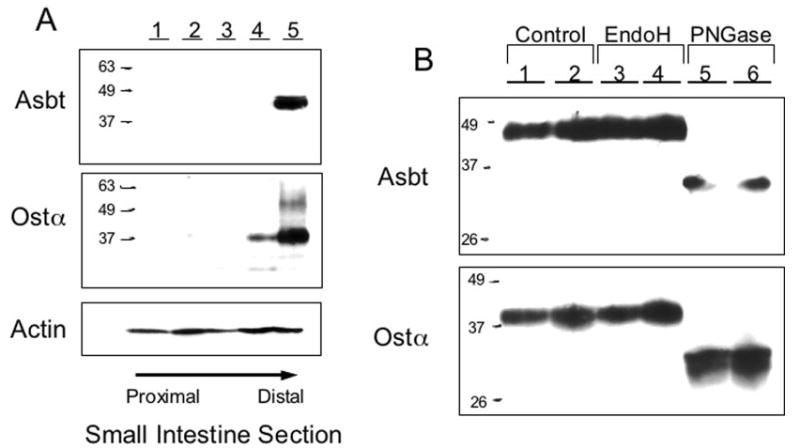
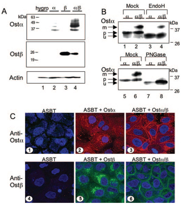
Bile acids are transported across the ileal enterocyte brush border membrane by the well characterized apical sodium-dependent bile acid transporter (Asbt) Slc10a2; however, the carrier(s) responsible for transporting bile acids across the ileocyte basolateral membrane into the portal circulation have not been fully identified. Transcriptional profiling of wild type and Slc10a2 null mice was employed to identify a new candidate basolateral bile acid carrier, the heteromeric organic solute transporter (Ost)alpha-Ostbeta. By Northern blot analysis, Ostalpha and Ostbeta mRNA was detected only in mouse kidney and intestine, mirroring the horizontal gradient of expression of Asbt in the gastrointestinal tract. Analysis of Ostalpha and Ostbeta protein expression by immunohistochemistry localized both subunits to the basolateral surface of the mouse ileal enterocyte. The transport properties of Ostalpha-Ostbeta were analyzed in stably transfected Madin-Darby canine kidney cells. Co-expression of mouse Ostalpha-Ostbeta, but not the individual subunits, stimulated Na(+)-independent bile acid uptake and the apical-to-basolateral transport of taurocholate. In contrast, basolateral-to-apical transport was not affected by Ostalpha-Ostbeta expression. Co-expression of Ostalpha and Ostbeta was required to convert the Ostalpha subunit to a mature glycosylated endoglycosidase H-resistant form, suggesting that co-expression facilitates the trafficking of Ostalpha through the Golgi apparatus. Immunolocalization studies showed that co-expression was necessary for plasma membrane expression of both Ostalpha and Ostbeta. These results demonstrate that the mouse Ostalpha-Ostbeta heteromeric transporter is a basolateral bile acid carrier and may be responsible for bile acid efflux in ileum and other ASBT-expressing tissues.


Figures

Fig. 1
Fig. 1. Expression of Ostα and Ostβ mRNA in wild type and Slc10a2 null mice
A, the small intestine (subdivided into five equal segments) was used to isolate total RNA. Ostα, Ostβ, Ibabp, and actin mRNA levels were then measured in pooled aliquots (5 male 129S6/SvEv mice, 4 months of age) of total RNA (10 μg) by Northern blot hybridization. B, pooled aliquots (3 female 129S6/SvEv mice, 7 months of age) of total RNA (10 μg) from cecum (Ce), proximal colon (Pc), and distal colon (Dc) were subjected to Northern blot hybridization using 32P-labeled Ostα, Ostβ, or cyclophilin probes.
Fig. 2
Fig. 2. Tissue distribution of mouse Ostα and Ostβ mRNA
A, tissue distribution of Ostα and Ostβ mRNA in the mouse. A blot containing 2 μg of poly(A)+ mRNA from the indicated mouse tissues was hybridized with a radiolabeled probe from the Ostα (upper panel) or Ostβ (lower panel) cDNA. Control experiments with a cyclophilin probe revealed that mRNA was present in all lanes of the blot. B, gastrointestinal tissue distribution of Asbt, Ostα, Ostβ, and Mrp3 mRNA in the mouse. A blot containing pooled aliquots of total RNA (10 μg) (3 female 129S6/SvEv mice, 7 months of age) from the indicated tissues was hybridized to the indicated radiolabeled probes for the Asbt, Ostα, Ostβ, Mrp3, or cyclophilin cDNAs. C, Asbt, Ostα, and Ostβ expression in the indicated mouse tissues was determined by real-time PCR. The threshold values (CT) are the means of triplicate determinations, and expression was normalized for GAPDH expression. The normalized threshold values are plotted as a percent of the ileal values (% of ileum). The threshold values determined in ileum were 19.4, 20.1, and 19.0 for Asbt, Ostα, and Ostβ, respectively.
Fig. 3
Fig. 3. Localization of Asbt and Ostα protein in the mouse small intestine and their sensitivity to glycosidase treatment
A, immunoblotting analysis of mouse small intestinal tissue segments. The small intestine was divided into five segments of equal length and used for membrane isolation. Aliquots of intestinal membrane protein (50 μg) were subjected to immunoblotting analysis using antibodies to the Asbt, Ostα, or β-actin, as indicated. B, glycosidase sensitivity of Asbt and Ostα. Aliquots of ileal membrane protein (50 μg) were incubated in the absence (lanes 1 and 2) or presence of Endo H (lanes 3 and 4) or PNGase F (lanes 5 and 6) and then subjected to immunoblotting analysis using the indicated antibodies.
Fig. 4
Fig. 4. Immunolocalization of Ostα and Ostβ in mouse ileum
Sections (6 μm) of mouse ileum were fixed with 3.7% formaldehyde/PBS, permeabilized with 0.05% Tween 20, and stained with polyclonal antibodies raised against Asbt (A and D), Ostα (B and E), or Ostβ (C and F) to determine their tissue and subcellular localization.
Fig. 5
Fig. 5. Expression, functional activity and glycosidase sensitivity of Ostα and Ostβ in HEK293 cells
A, on day 0, HEK293 cells were seeded in 35-mm dishes. On day 1, the cells were transfected with β-galactosidase, Ostα, Ostβ, or Ostα-Ostβ. On day 2, the cells were washed and incubated in Hanks’ buffered saline containing 25 μm [3H]taurocholate for 30 min at 37 °C. Cells were then washed and lysed to determine cell-associated radioactivity and protein. The uptake is expressed as pmol of taurocholate transported/mg of protein (mean ± S.D., n = 3) and is corrected for the background uptake in β-galactosidase transfected cells (15.6 ± 0.6). Taurocholate uptake was significantly increased following co-transfection with Ostα plus Ostβ (p < 0.001). B, on day 0, HEK293 cells were seeded in 60-mm dishes. On day 1, the cells were transfected with Ostα, Ostβ, or Ostα-Ostβ. On day 2, the cells were lysed, and 100 μg of cell protein was incubated in the absence (lanes 1, 2, 7, 8) or presence of Endo H (lanes 3, 4, 9, 10), or PNGase F (lanes 5, 6, 11, 12). Samples were then subjected to immunoblotting analysis for Ostα (lanes 1–6) and Ostβ (lanes 7–12). The migration of the precursor (p), mature glycosylated (m), and unglycosylated (u) forms are indicated. C, on day 0, HEK293 cells were seeded onto glass coverslips. On day 1, the cells were transfected with Ostα (panels 1 and 4), Ostβ (panels 2 and 5), or Ostα-Ostβ (panels 3 and 6). After 24 h, the cells were fixed with 3.7% formaldehyde/PBS and permeabilized using a solution of 1% bovine serum albumin and 0.1% saponin in PBS. The cells were then stained with mouse M2 anti-FLAG to detect Ostα (red), rabbit polyclonal anti-Ostβ (green), and To-Pro-3 (blue) to visualize nuclei and then viewed using laser scanning confocal microscopy.
Fig. 6
Fig. 6. Expression and glycosidase sensitivity of Ostα and Ostβ in stably transfected MDCK cells
A, MDCK-ASBT cells stably transfected with the indicated plasmids were lysed, and 100 μg of cell protein was subjected to immunoblotting analysis for Ostα, Ostβ, and β-actin, as indicated. B, MDCK-ASBT cells stably transfected with Ostα (α) or Ostα-Ostβ (α/β) were lysed, and 100 μg of cell protein was incubated in the absence (lanes 1, 2, 5, 6) or presence (lanes 3, 4) of Endo H or PNGase F (lanes 7, 8). Samples were then subjected to immunoblotting analysis for Ostα. C, MDCK-ASBT cells stably transfected with pHygro (panels 1 and 4), Ostα (panels 2 and 5), or OstαIRESOstβ (panels 3 and 6) were seeded onto glass coverslips. After 24 h, the cells were fixed with 3.7% formaldehyde/PBS and permeabilized using a solution of 1% bovine serum albumin and 0.1% saponin in PBS. The cells were then stained with rabbit polyclonal anti-Ostα (red, panels 1–3), rabbit polyclonal anti-Ostβ (green, panels 4–6) and To-Pro-3 (blue) to detect nuclei and then viewed using laser scanning confocal microscopy.
Fig. 7
Fig. 7. Bile acid uptake activity in stably transfected MDCK cells
On day 0, MDCK cells stably transfected with the indicated plasmids were seeded onto 24-well plates. On day 2, expression of the transfected plasmids was induced by the addition of 10 mm sodium butyrate. After 20 h, the cell monolayers were washed and incubated for 30 min at 37 °C with 10 μm [3H]taurocholate either in the presence of 137 mm Na+ (A) or K+ (B). The cells were then washed and processed to determine cell-associated protein and radioactivity. Uptake values (mean ± S.D.) are expressed as pmol of taurocholate/mg of cell protein.
Fig. 8
Fig. 8. Trans-cellular transport of taurocholate in stably transfected MDCK cells
MDCK cells transfected with the indicated plasmids were plated onto Transwell filter inserts on day 0. Expression of the transfected plasmids was induced by the addition of 10 mm sodium butyrate on day 7. On day 8, either the apical or the basolateral Transwell chamber received 10 μm [3H]taurocholate, and aliquots of media in the opposite chamber were sampled over 60 min. Data for the apical to basolateral transport are shown in the line graph. Data for basolateral to apical transport after 60 min are shown in the inset. Values are means ± S.D., n = 3.

References

    1. Shneider, B. L. (2001) J. Pediatr. Gastroenterol. Nutr.32, 407–417 - PubMed
    1. Oelkers, P., Kirby, L. C., Heubi, J. E., and Dawson, P. A. (1997) J. Clin. Investig.99, 1880–1887 - PMC - PubMed
    1. Dawson, P. A., Haywood, J., Craddock, A. L., Wilson, M., Tietjen, M., Kluckman, K., Maeda, N., Parks, J. S. (2003) J. Biol. Chem.278, 33920–33927 - PubMed
    1. Weinberg, S. L., Burckhardt, G., Wilson, F. A. (1986) J. Clin. Investig.78, 44–50 - PMC - PubMed
    1. Lin, M. C., Weinberg, S. L., Kramer, W., Burckhardt, G., and Wilson, F. A. (1988) J. Membr. Biol.106, 1–11 - PubMed

Publication types