. 2005 Jan;128(Pt 1):174-88.
doi: 10.1093/brain/awh337. Epub 2004 Nov 24.

Progression from frontal-parietal to mesial-temporal epilepsy after fluid percussion injury in the rat

Affiliations

Progression from frontal-parietal to mesial-temporal epilepsy after fluid percussion injury in the rat

Raimondo D'Ambrosio et al. Brain. 2005 Jan.

Abstract

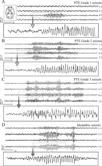
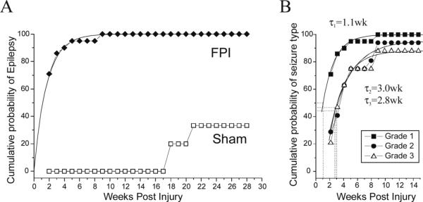
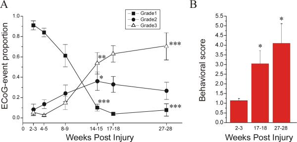
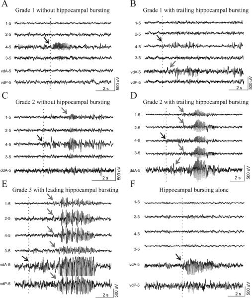
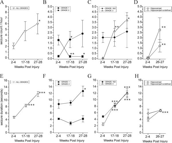
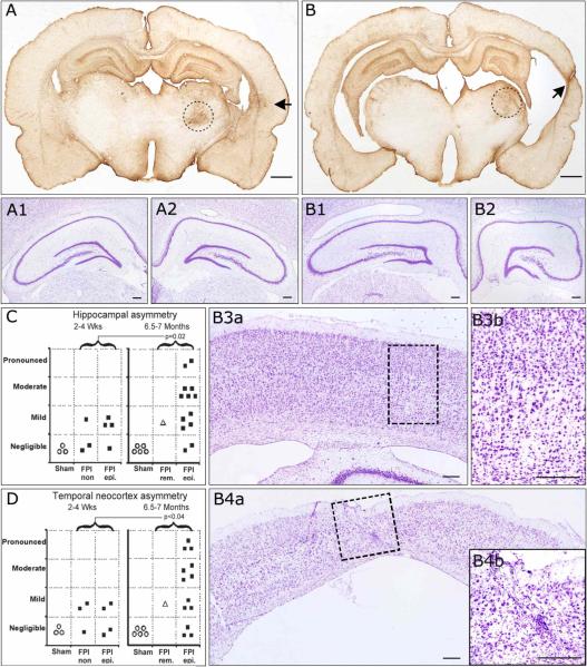
We recently described an in vivo model of post-traumatic epilepsy (PTE) in the rat where chronic spontaneous recurrent seizures appear following a single episode of fluid percussion injury (FPI). PTE, studied during the first 2 months post-injury, was focal and seizures originated predominantly from the frontal-parietal neocortex at or around the injury site. However, rarer bilateral seizures originating from a different and undefined focus were also observed. To shed light on the Posttraumatic Epileptogenic mechanisms and on the generation of bilateral seizures, we studied rats up to 7 months post-injury. In vivo paired epidural and depth-electrode recordings indicated that the anterior hippocampus evolves into an epileptic focus which initiates bilateral seizures. The rate of frontal-parietal seizures remained constant over time after 2 weeks post-injury, while the rate of hippocampal seizures greatly increased over time, suggesting that different mechanisms mediate neocortical and hippocampal post-traumatic epileptogenesis. Because of different temporal evolution of these foci, the epileptic syndrome was characterized by predominant frontal-parietal seizures early after injury, but by predominant mesio-temporal seizures at later time points. Pathological analysis demonstrated progressive hippocampal and temporal cortex pathology that paralleled the increase in frequency and duration of bilateral seizures. These results demonstrate that FPI-induced frontal-parietal epilepsy (FPE) progresses to mesial-temporal lobe epilepsy (MTLE) with dual pathology. These observations establish numerous similarities between FPI-induced and human PTE and further validate it as a clinically relevant model of PTE.


Figures

Figure 1
Figure 1. Different types of chronic recurrent seizures as revealed by ECoG following FPI
Inset top, left) Schematic of the locations of the five cortical electrodes (filled circles) and of the injury site (hollow circle) in respect to the rat skull. All panels represent continuous ECoG monitoring at 7 months post-FPI in wake animals. A) A representative grade 1 posttraumatic seizure detected exclusively by the peri-lesion electrode. B) A representative grade 2 posttraumatic seizure first detected by the peri-lesion electrode and then by multiple channels. C) A representative grade 3 posttraumatic seizure detected simultaneously by multiple channels. D) A representative idiopathic seizure, detected bilaterally and characterized by larger occipital epileptic discharge. These idiopathic seizures were absent until 5.5 months of age and represented 3.6% of all cortical discharge of FPI animals at 8 months of age. ECoG calibration bars are on the left of each panel. Dotted boxes highlight the portion of ECoG shown at higher temporal resolution in the rectangle underneath each panel. The numbers next to each ECoG trace indicate the electrodes by which the trace is recorded, and its reference.
Figure 2
Figure 2. Probability of unprovoked seizures following severe lateral FPI
A) The cumulative probability of detecting unprovoked epileptiform ECoG events in post-FPI (filled circles) and sham-injured (hollow squares) rats is plotted versus time after injury. The incidence of unprovoked seizures in the sham-injured CD Sprague-Dawley rats used for this study is zero up to 4.5 months post-surgery (5.5 months old). B) The cumulative probability of developing grade 1 (filled squares), grade 2 (filled circles), and grade 3 (hollow triangles) seizures by chronic ECoG is plotted versus time after injury. The temporal increase in probability of developing each seizure type had half time τ1=1.1 weeks for grade 1 seizures, τ2=3.0 weeks for grade 2, and τ3=2.8 weeks for grade 3 seizures.
Figure 3
Figure 3. Temporal evolution of the FPI-induced posttraumatic epileptic syndrome
Electrical and behavioral correlates of PTE progression are assessed in 8 epileptic animals. A) The proportions of ECoG events of grade 1, 2, and 3 are plotted over time from 2 to 28 weeks post-injury. Focal frontal-parietal seizures (grade 1; filled square) represented the most common seizure type 2−3 weeks post-injury, but then progressively decreased in proportion over time. Focal neocortical spreading seizures (grade 2; filled circles) were rare at 2−3 weeks post-injury, but then increased in proportion over time and peaked at 14−15 weeks post-injury. Seizures that did not originate from the frontal-parietal cortex and appeared bilateral in onset (grade 3; hollow triangle) were rare up to 8−9 weeks post-injury, and then increased up to 27−28 weeks post-injury. Note the overall increase in seizures’ bilateral spread over time post-injury (grades 2&3 combined). B) The behavioral score during epileptiform ECoG events is plotted versus time post-injury. Note the progression of the severity of behavioral seizures over time post-injury. Data are presented as Mean±S.E.M. Statistics with paired t-test (* p<0.05; ** P≤0.01; *** P≤0.001, vs 2−3 weeks).
Figure 4
Figure 4. Independent firing and crosstalk of the frontal-parietal neocortical and antero-hippocampal epileptic foci
Paired epidural and depth-electrode recordings performed in 6 epileptic animals 6.5−7 months post-injury. A) A grade 1 seizure detected by the peri-lesion epidural electrode does not recruit the hippocampus, indicating the existence and independent firing of a focus in the frontal-parietal neocortex. B) Epileptic activity is first detected by the peri-lesion epidural electrode during a grade 1 seizure, and then in the anterior hippocampus, indicating the neocortical focus recruited the hippocampus. C) The hippocampus shows no epileptiform activity during the occurrence of a grade 2 seizure that originated around the injury site and then propagated to the frontal contralateral cortex. D) Epileptic activity is first detected by the perilesional epidural electrode in the frontal-parietal cortex, and then spreads ipsi- and contralaterally, during a grade 2 seizure, and to the anterior hippocampus, indicating the neocortical focus recruited the hippocampus. E) Epileptiform activity is first detected in the anterior hippocampus and then simultaneously in the ipsi- and contralateral neocortex, indicating that grade 3 seizures originate in the hippocampus. F) Epileptiform activity is detected only in the hippocampus, in absence of any neocortical discharge, indicating the existence and independent firing of an epileptic focus in the hippocampus. Scale bars apply to all four traces in each panel. Epileptic bursts shown in E and F are cut off at ±500μV by gain saturation. Dotted lines and black arrows indicate the beginning of epileptiform activity. Gray arrows indicate propagated epileptic activity. vdA = vertical depth electrode placed in the anterior hippocampus; vdP = vertical depth electrode placed in the posterior hippocampus; ddA = diagonal depth electrode placed in the anterior hippocampus. The text to the left of each ECoG trace indicate the electrode by which the trace is recorded and its reference.
Figure 5
Figure 5. Temporal evolution of rate of occurrence and duration of cortical and hippocampal discharge
Cortical and hippocampal discharge rate of occurrence (A-D) and duration (E-H) are shown from 2 to 28 weeks post-injury. Cortical discharge occurring during all seizure grades (1, 2, and 3) increased in frequency over time (A). Grade 1 seizures decreased, while grade 2 seizures increased in frequency over time (B). The rate of seizures originating from the frontal-parietal neocortex (grade 1 and 2 combined) did not change from 2−4 weeks to 27−28 weeks post-injury, while the rate of seizures not originating from that focus (grade 3) dramatically increased over time post-injury (C). The frequency of hippocampal seizures increased over time post-injury. Because seizures detected simultaneously in both hippocampus and cortex may also be originating from the hippocampus, the frequency of hippocampal+undefined seizures is shown (D). Cortical discharge duration (all seizure grades) increased over time post-injury (E). The duration of grade 1 seizures did not significantly increase over time, while grade 2 seizures did (F). The duration of seizures originating from the frontal-parietal focus (grades 1 and 2) increased, as did the duration of grade 3 seizures that do not originate from it (G). We did not detect a significant increase over time post-injury in the duration of hippocampal discharge during hippocampal seizures. However, the duration of hippocampal+undefined seizures significantly increased over time post-injury (H). Data are presented as mean±SEM. Statistical comparisons were performed with Wilcoxon Signed Rank test (A-C) and With Mann-Whitney U test (D-H) vs 2−4 wks time point (* p<0.05; ** P≤0.01; *** P≤0.001).
Figure 6
Figure 6. Progressive hippocampal and temporal cortex pathology in posttraumatic epileptic rat
Coronal sections obtained from bregma −4 mm through bregma −5 mm and stained for cresyl violet and GFAP. A) GFAP immunoreactivity of an epileptic animal 7 months post-injury demonstrating no hippocampal and mild temporal cortex asymmetry. At higher magnification, cresyl violet staining shows symmetric contralateral (A1) and ipsilateral (A2) hippocampi and no loss of laminar features. B) GFAP immunoreactivity of another epileptic animal 7 months post-injury demonstrating pronounced hippocampal and temporal cortex asymmetry. At higher magnification, cresyl violet staining shows asymmetric contralateral (B1) and ipsilateral (B2) hippocampi, with evident atrophy of ipsilateral CA3 and CA1subregions. The contralateral temporal cortex showed no atrophy (B3a) and normal laminar structure (B3b). However, the ipsilateral temporal cortex was atrophic (B4a) and showed pronounced loss of neurons and laminar features, and increased small nuclei representing reactive glia (B4b), all typically associated with temporal cortex sclerosis. Hippocampal (C) and temporal cortex (D) asymmetry increased over time post-injury in the population of FPI animals. Statistics with one-tailed Mann-Whitney U test. FPI rem. = case of PTE remission following FPI. FPI epi. = persistently epileptic animals. FPI n.epi. = not epileptic animals. Scale bars: 1mm for A and B; 250 μm for A1−2 and B1-B4b. Black arrows in A and B indicate temporal foci of glial reactivity. Dotted circles in A and B delineate the thalamic injury present in all rpFPI animals. Dotted rectangles in B3a and B4a delineate the areas magnified in B3b and B4b, respectively.

Similar articles

Cited by

References

    1. Annegers JF, Rocca WA, Hauser WA. Causes of epilepsy: contributions of the Rochester epidemiology project. Mayo Clin Proc. 1996;71:570–575. - PubMed
    1. Annegers JF, Hauser WA, Coan SP, et al. A population-based study of seizures after traumatic brain injuries. N Engl J Med. 1998;338:20–24. - PubMed
    1. Arruda F, Cendes F, Andermann F, Dubeau F, Villemure JG, Jones-Gotman M, Poulin N, Arnold DL, Olivier A. Mesial atrophy and outcome after amygdalohippocampectomy or temporal lobe removal. Ann Neurol. 1996;40(3):446–50. - PubMed
    1. Benbadis SR, Heriaud L, Tatum WO, Vale FL. Epilepsy surgery, delays and referral patterns-are all your epilepsy patients controlled? Seizure. 2003;12(3):167–70. - PubMed
    1. Bertram EH, Zhang DX. Thalamic excitation of hippocampal CA1 neurons: a comparison with the effects of CA3 stimulation. Neuroscience. 1999;92(1):15–26. - PubMed

Publication types

MeSH terms