. 2004 Dec 7;101(49):16990-5.
doi: 10.1073/pnas.0407402101. Epub 2004 Nov 24.

Coordinated assembly of human translation initiation complexes by the hepatitis C virus internal ribosome entry site RNA

Affiliations

Coordinated assembly of human translation initiation complexes by the hepatitis C virus internal ribosome entry site RNA

Hong Ji et al. Proc Natl Acad Sci U S A. .

Abstract

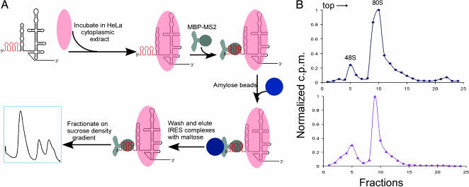
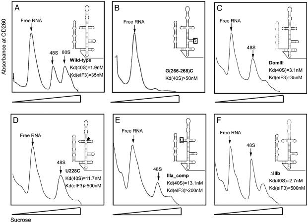
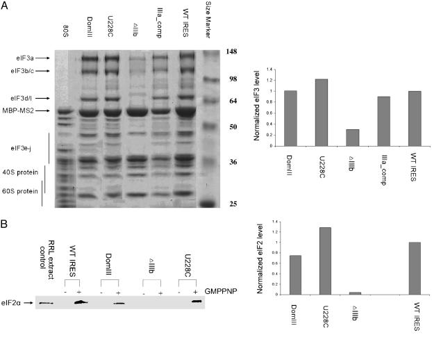
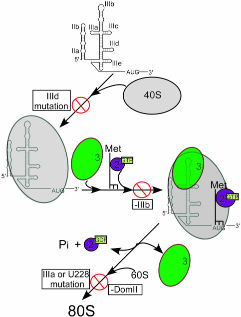
Protein synthesis in all cells begins with recruitment of the small ribosomal subunit to the initiation codon in a messenger RNA. In some eukaryotic viruses, RNA upstream of the coding region forms an internal ribosome entry site (IRES) that directly binds to the 40S ribosomal subunit and enables translation initiation in the absence of many canonical translation initiation factors. The hepatitis C virus (HCV) IRES RNA requires just two initiation factors, eukaryotic initiation factor (eIF) 2 and eIF3, to form preinitiation 48S ribosomal complexes that subsequently assemble into translation-competent ribosomes. Using an RNA-based affinity purification approach, we show here that HCV IRES RNA facilitates eIF2 function through its interactions with eIF3 and the 40S ribosomal subunit. Although the wild-type IRES assembles normally into 48S and 80S ribosomal complexes in human cell extract, mutant IRES RNAs become trapped at the 48S assembly stage. Trapped 48S complexes formed by IRES mutants with reduced eIF3 binding affinity nonetheless contain eIF3, consistent with inherent eIF3-40S subunit affinity. Intriguingly, however, one of these IRES mutants prevents stable association of both eIF3 and eIF2, preventing initiator tRNA deposition and explaining the block in 80S assembly. In contrast, an IRES mutant unable to induce a conformational change in the 40S subunit, as observed previously by single-particle cryoelectron microscopy, blocks 80S formation at a later stage in assembly. These data suggest that the IRES RNA coordinates interactions of eIF3 and eIF2 on the ribosome required to position the initiator tRNA on the mRNA in the ribosomal peptidyl-tRNA site (P site).


Figures

Fig. 1.
Fig. 1.
IRES-containing ribosomal complex purification. (A) Strategy for affinity purification of IRES-containing complexes from HeLa cell extract. HCV IRES RNAs containing three MS2 recognition hairpins at the 5′ end (shown in red) were incubated in HeLa cell cytoplasmic extract; IRES-bound complexes were affinity-purified by binding to a chimeric MBP-MS2 fusion protein and isolating by amylose affinity chromatography (see Materials and Methods). Translation initiation complexes were then separated and purified by sucrose density gradient centrifugation. (B) Ribosomal complex assembly in HeLa cell lysate using 32P-end-labeled wild-type IRES (Upper) or 32P-end-labeled MS2-tagged wild-type IRES (Lower). Ribosomal complexes were fractionated by sucrose density gradient centrifugation, and radioactivity in each fraction was determined by PhosphorImager analysis (see Materials and Methods), with values normalized by dividing by the maximum cpm value observed; sedimentation was from left to right.
Fig. 2.
Fig. 2.
Sucrose density gradient analysis of translation complexes bound by wild-type and mutant forms of the HCV IRES. Affinity-tagged IRES RNAs were incubated in HeLa cell extract at a concentration of extract yielding half-maximal binding of wild-type IRES. Plots of absorbance at 260 nm versus sucrose density are shown; peaks corresponding to free RNA, 48S, and 80S are indicated (verified by denaturing PAGE and electrospray mass spectrometry). (A) Wild-type IRES. (BF) Translation-defective IRES constructs containing the following mutations and translation complex binding defects relative to the wild-type HCV IRES (12, 23). (B) G(266–268)C mutation in the IIId loop, >25-fold-reduced 40S binding affinity. (C) DomIII, no change in 40S or eIF3 binding affinities. (D) U228C, >15-fold-reduced eIF3 binding affinity. (E) IIIa_Comp, >6-fold-reduced eIF3 binding affinity. (F) ΔIIIb, >15-fold-reduced eIF3 binding affinity.
Fig. 3.
Fig. 3.
Analysis of initiation factor presence within translation complexes assembled on wild-type and mutant HCV IRES RNAs. (A Left) Coomassie blue-stained 10% SDS/PAGE gel of affinity-purified 48S and 80S samples; IRES constructs are indicated at the top, protein size markers are labeled at right, and bands corresponding to eIF3, 40S, and 60S proteins are indicated at left. The identities of eIF3 bands were verified by Western blotting (data not shown). (Right) Relative eIF3 levels in wild-type IRES and mutant IRES-bound complexes. For each sample, three eIF3 bands were quantitated by using imagej software, and the summed intensities were divided by the intensity of the MBP-MS2 band as a loading control; all values were normalized to that of the wild-type IRES-containing sample. Each value shown is the average of two independent experiments. (B Left) Western blot analysis of 48S complexes in the absence and presence of the nonhydrolyzable GTP analog GMPPNP using an anti-eIF2α antibody. Equal amounts of sample based on OD260 were applied to each lane. (Right) Relative eIF2 levels in wild-type and mutant IRES-bound complexes. Band intensities were quantitated by using imagej software and divided by the intensity of ribosomal protein bands in each lane to control for sample loading differences; all values were normalized to that of the wild-type IRES-containing sample. Each value shown, except for U228C, is the average of two independent experiments.
Fig. 4.
Fig. 4.
Model for HCV IRES-coordinated assembly of human translation initiation complexes. 40S subunit binding to IRES RNA with an intact IIId loop is required for IRES-40S association; eIF3 interaction with the IIIb region of the IRES is required for stable association of eIF3 and eIF2 in the 48S complex, whereas eIF3 contacts to the junction of stems IIIa, -b and -c, as well as the 40S conformational change induced by DomII, are necessary for downstream events required for 60S subunit joining.

Comment in

  • Biography of Jennifer A. Doudna.
    Marino M. Marino M. Proc Natl Acad Sci U S A. 2004 Dec 7;101(49):16987-9. doi: 10.1073/pnas.0408147101. Epub 2004 Dec 1. Proc Natl Acad Sci U S A. 2004. PMID: 15574498 Free PMC article. No abstract available.

Similar articles

Cited by

References

    1. Kozak, M. (1989) J. Cell Biol. 108, 229–241. - PMC - PubMed
    1. Hershey, J. W. B. & Merrick, W. C. (2000) in Translational Control of Gene Expression, eds. Sonenberg, N., Hershey, J. W. B. & Mathews, M. B. (Cold Spring Harbor Lab. Press, Plainview, NY), Vol. 39, pp. 33–88.
    1. Pestova, T. V., Kolupaeva, V. G., Lomakin, I. B., Pilipenko, E. V., Shatsky, I. N., Agol, V. I. & Hellen, C. U. (2001) Proc. Natl. Acad. Sci. USA 98, 7029–7036. - PMC - PubMed
    1. Hellen, C. U. & Sarnow, P. (2001) Genes Dev. 15, 1593–1612. - PubMed
    1. Merrick, W. C. (2004) Gene 332, 1–11. - PubMed

Publication types

MeSH terms

Substances