. 2004 Dec 7;101(49):17216-21.
doi: 10.1073/pnas.0404761101. Epub 2004 Nov 24.

Liver-targeted disruption of Apc in mice activates beta-catenin signaling and leads to hepatocellular carcinomas

Affiliations

Liver-targeted disruption of Apc in mice activates beta-catenin signaling and leads to hepatocellular carcinomas

S Colnot et al. Proc Natl Acad Sci U S A. .

Abstract

Although inappropriate activation of the Wnt/beta-catenin pathway has been implicated in the development of hepatocellular carcinoma (HCC), the role of this signaling in liver carcinogenesis remains unclear. To investigate this issue, we constructed a mutant mouse strain, Apc(lox/lox), in which exon 14 of the tumor-suppressor gene adenomatous polyposis coli (Apc) is flanked by loxP sequences. i.v. injection of adenovirus encoding Cre recombinase (AdCre) at high multiplicity [10(9) plaque-forming units (pfu) per mouse] inactivated the Apc gene in the liver and resulted in marked hepatomegaly, hepatocyte hyperplasia, and rapid mortality. beta-Catenin signaling activation was demonstrated by nuclear and cytoplasmic accumulation of beta-catenin in the hepatocytes and by the induction of beta-catenin target genes (glutamine synthetase, glutamate transporter 1, ornithine aminotransferase, and leukocyte cell-derived chemotaxin 2) in the liver. To test a long-term oncogenic effect, we inoculated mice with lower doses of AdCre (0.5 x 10(9) pfu per mouse), compatible with both survival and persistence of beta-catenin-activated cells. In these conditions, 67% of mice developed HCC. beta-Catenin signaling was strongly activated in these Apc-inactivated HCCs. The HCCs were well, moderately, or poorly differentiated. Indeed, their histological and molecular features mimicked human HCC. Thus, deletion of Apc in the liver provides a valuable model of human HCC, and, in this model, activation of the Wnt/beta-catenin pathway by invalidation of Apc is required for liver tumorigenesis.


Figures

Fig. 1.
Fig. 1.
Activation of β-catenin signaling, hepatomegaly, and altered survival in Apc-inactivated mice infected with AdCre at high multiplicity. (A) Various alleles of Apc in floxed mice. Restriction sites: E, EcoRI; B, BamHI; X, XbaI; EV, EcoRV. Exons 11–15 are represented by black bars; primers and the lengths of the PCR fragments generated are indicated by arrows. (B) Cre delivery strategy. (C) Survival curve for Apclox/lox and Apc+/lox mice after injection of various doses of AdCre. (D) Liver as a percentage of body weight and proliferation quantified by Ki67 scores in Apclox/lox compared with Apc+/lox Cre-infected livers. *, P < 0.001; **, P = 0.0028. (E) PCR of DNA extracted from livers of Apc floxed mice showing the accumulation of the ApcΔex14 band in AdCre-treated livers compared with noninjected (NI) mouse livers. (F) β-Catenin immunostaining of membranes in Apc+/lox injected mice (Apc+/–). (G) Cytosolic and nuclear accumulation of β-catenin staining in injected, Apclox/lox mice (Apc–/–). (Scale bars in F and G:20 μm.)
Fig. 2.
Fig. 2.
Dose-dependent Apc inactivation and cell-autonomous induction of liver-specific β-catenin target genes. (A) β-Catenin and GS immunostaining of Apc+/– and Apc–/– mice injected with various doses of AdCre. (B and C) Coimmunostaining of β-catenin and its liver-specific targets (GS, GLT1, and LECT2, in blue) in perivenous (B) and nonperivenous (C) areas. β-Catenin stains red-brown at the membrane of every hepatocyte and accumulates in the cytosol and nuclei of Apc–/– hepatocytes. CV, centrolobular vein; PS, portal space. (Scale bars: A, 100 μm; B and C, 20 μm.) (D) Percentage of hepatocytes that are GS+ in nonperivenous areas of Apc–/– mice, according to dose of AdCre. Filled ovals represent mice with a hepatomegaly (liver > 8.5% of body weight), and open ovals represent mice without hepatomegaly.
Fig. 3.
Fig. 3.
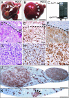
Hepatocarcinogenesis from Apc–/– hepatocytes. (A and B) HCC1 and HCC2 developed 8 and 9 months, respectively, after AdCre infection. (C) PCR analysis from tumoral DNA shows the loss of the Apclox allele and the gain of the ApcΔex14 allele. (DF) MD HCCs. (GI) PD HCCs. Hematoxylin/eosin staining shows atypical hepatocytes organized in a trabecular (D) or pseudoglandular (G) pattern; β-catenin mislocalizes in the nucleus (E and H), and GS is overexpressed (F and I). Nontumoral (NT) and tumoral (T) tissues are delineated by a dashed line. (J and K) GS immunostaining in macronodular HCCs (J) and in a micronodular preneoplastic lesion (K), indicated by arrows. (Scale bars: A and B, 1 cm; DI, 50 μm; J and K, 1 mm.)
Fig. 4.
Fig. 4.
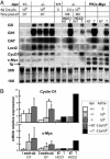
Comparative expressions of β-catenin-dependent target genes in Apc–/– hyperplastic livers and in β-catenin-activated HCC. (A) Northern blot analysis of livers from adult (3–5 months old) Apc floxed mice, not injected (0) or on D7 after injection of 109 pfu of AdCre (109). Apc–/– and PK/c-Myc tumoral (T) or nontumoral (NT) livers are also shown. HCC1 and HCC2 are the WD and MD HCCs shown in Fig. 3 A and B, respectively. Note for c-Myc the presence of two bands in PK/c-Myc livers: The lower band is the c-Myc transgene (tg) mRNA, and the upper band is the endogenous c-Myc mRNA. OAT, ornithine aminotransferase; 28S and 18S are the rRNAs. (B) Real-time quantitative RT-PCR analysis of cyclin D1 (CycD1) and c-Myc mRNAs extracted from D7 infected livers, HCC1 and HCC2. For analyses at D7, three 1-month-old and four 5-month-old animals were analyzed in each group. No significant differences were observed, except those marked by an asterisk, where P =0.016.
Fig. 5.
Fig. 5.
p53 immunostaining of Apc–/– HCC. (A) p53 accumulation in a hyperplastic liver of large T antigen-expressing ATIII-SV40 transgenic mice (37), used as a positive control for nuclear staining. (B) No p53 staining was seen in an Apc–/– MD HCC. (C) Intense p53 staining was seen in the nuclei of tumoral cells from an Apc–/– PD HCC. (Scale bars: 50 μm; 20 μmin Insets.)

Similar articles

Cited by

References

    1. Befeler, A. S. & Di Bisceglie, A. M. (2002) Gastroenterology 122, 1609–1619. - PubMed
    1. Buendia, M. A. (2000) Semin. Cancer Biol. 10, 185–200. - PubMed
    1. Giles, R. H., van Es, J. H. & Clevers, H. (2003) Biochim. Biophys. Acta 1653, 1–24. - PubMed
    1. de La Coste, A., Romagnolo, B., Billuart, P., Renard, C. A., Buendia, M. A., Soubrane, O., Fabre, M., Chelly, J., Beldjord, C., Kahn, A. & Perret, C. (1998) Proc. Natl. Acad. Sci. USA 95, 8847–8851. - PMC - PubMed
    1. Chen, T. C., Hsieh, L. L., Ng, K. F., Jeng, L. B. & Chen, M. F. (1998) Cancer Lett. 134, 23–28. - PubMed

Publication types