. 2005 Feb;16(2):757-68.
doi: 10.1091/mbc.e04-08-0672. Epub 2004 Nov 24.

Fibronectin matrix turnover occurs through a caveolin-1-dependent process

Affiliations

Fibronectin matrix turnover occurs through a caveolin-1-dependent process

Jane Sottile et al. Mol Biol Cell. 2005 Feb.

Abstract

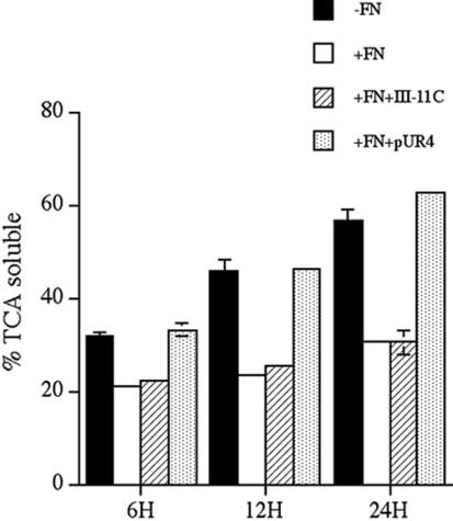
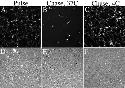
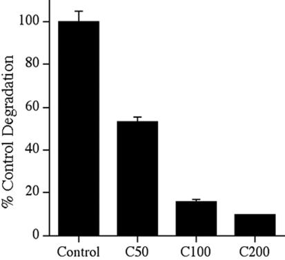
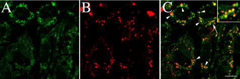
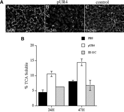
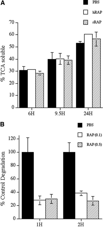
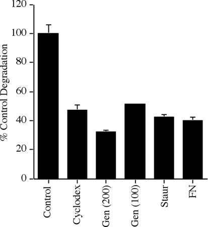
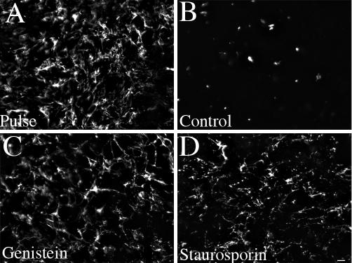
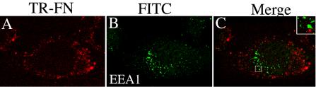
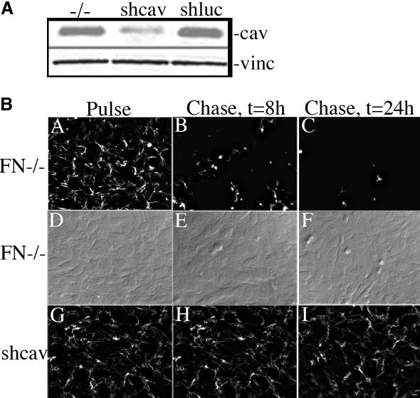
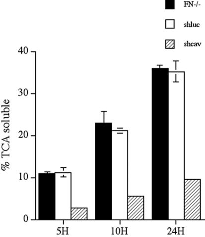
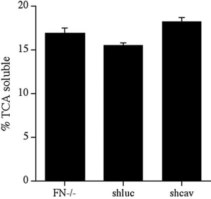
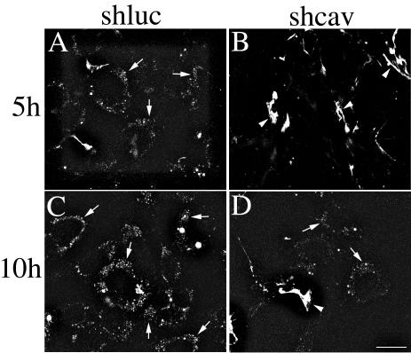
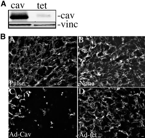
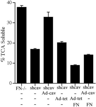
Extracellular matrix remodeling occurs during development, tissue repair, and in a number of pathologies, including fibrotic disorders, hypertension, and atherosclerosis. Extracellular matrix remodeling involves the complex interplay between extracellular matrix synthesis, deposition, and degradation. Factors that control these processes are likely to play key roles in regulating physiological and pathological extracellular matrix remodeling. Our data show that fibronectin polymerization into the extracellular matrix regulates the deposition and stability of other extracellular matrix proteins, including collagen I and thrombospondin-1 (Sottile and Hocking, 2002. Mol. Biol. Cell 13, 3546). In the absence of continual fibronectin polymerization, there is a loss of fibronectin matrix fibrils, and increased levels of fibronectin degradation. Fibronectin degradation occurs intracellularly after endocytosis and can be inhibited by chloroquine, an inhibitor of lysosomal degradation, and by caveolae-disrupting agents. Down-regulation of caveolin-1 by RNAi inhibits loss of fibronectin matrix fibrils, fibronectin internalization, and fibronectin degradation; these processes can be restored by reexpression of caveolin-1. These data show that fibronectin matrix turnover occurs through a caveolin-1-dependent process. Caveolin-1 regulation of fibronectin matrix turnover is a novel mechanism regulating extracellular matrix remodeling.


Figures

Figure 1.
Figure 1.
Fibronectin degradation is inhibited by fibronectin polymerization. FN-null MF were incubated overnight with 125I-labeled fibronectin. Cells were washed and then incubated for the indicated time with culture medium containing (+FN) or lacking unlabeled fibronectin (-FN) and containing or lacking pUR4 or the control his-tagged peptide, III-11C for the indicated times. The cell culture supernatant was collected and precipitated with TCA as described in Materials and Methods. The amount TCA soluble counts is expressed as a percentage of the total counts present in each well. Error bars represent the range of duplicate determinations.
Figure 2.
Figure 2.
Fibronectin matrix turnover does not occur at 4°C. FN-null MF were incubated overnight with 20 nM FITC-conjugated fibronectin. Cell were then either processed for immunofluorescence (Pulse, A) or were washed, and then incubated with culture medium lacking fibronectin (Chase) at 37°C (B) or 4°C (C) for 8 h. Cells were fixed and permeabilized. FITC-fibronectin staining (A–C) and the corresponding phase images (D–F) are shown. Bar, 10 μm.
Figure 3.
Figure 3.
Fibronectin degradation is inhibited by chloroquine. FN-null MF were incubated overnight with 125I-labeled fibronectin. Cells were washed and then incubated with culture medium lacking fibronectin and lacking (control) or containing 50–200 μM chloroquine (C50, C100, C200) for 10 h. The cell culture supernatant was collected, and precipitated with TCA as described in the legend to Figure 1. The amount of degradation that occurred in the absence of chloroquine was set equal to 100%. Error bars represent the range of duplicate determinations.
Figure 4.
Figure 4.
Colocalization of fibronectin with the lysosomal marker, LAMP-1. FN-null MF were incubated with Texas Red–conjugated fibronectin (20 nM) overnight. Cells were washed and then incubated for 25 h in cell culture media lacking fibronectin, but containing 50 μM chloroquine. Chloroquine was used to inhibit lysosomal degradation and enhance the levels of intracellular fibronectin. Cells were then fixed, permeabilized, and incubated with an antibody to LAMP-1. Localization of Texas-Red-fibronectin (B) and LAMP-1 (A) are shown. (C) An overlay of the fibronectin and LAMP-1 images shown in A and B. Some areas of colocalization (arrowheads) of fibronectin and LAMP-1 are shown in yellow. It should be noted that yellow will only be seen if the intensity of the red and green fluors are approximately equal. The area marked by the arrow in C was magnified and is shown in the inset. The images are optical sections collected from a confocal z-series scan obtained with an Olympus confocal microscope. Bar, 10 μm.
Figure 5.
Figure 5.
(A) Fibronectin matrix stability in SMCs. Human aortic SMCs (passage 4) were seeded in serum-containing medium and grown to confluence. SMCs were then incubated with 250 nM pUR4 (B) or 250 nM control peptide, III-11C (C) for 24 h. At the time of protein addition, some cells were processed for immunofluorescence (A) to determine the amount of matrix deposited by the cells before pUR4 addition (t = 0). Cells were permeabilized and then incubated with a polyclonal antibody to fibronectin, followed by a FITC anti-rabbit antibody. Bar, 20 μm. (B) Fibronectin degradation in SMC. Confluent cultures of rat SMCs were incubated overnight with 125I-fibronectin. Cells were washed and then incubated with culture medium lacking (PBS) or containing 250 nM pUR4 or the control III-11C peptide for the indicated times. The amount of TCA-soluble counts was determined as described in Materials and Methods and is expressed as a percentage of the total counts present in each well. Error bars represent the average of duplicate samples, and the error bars the range.
Figure 6.
Figure 6.
(A) Fibronectin degradation is not inhibited by RAP. FN-null MF were incubated overnight with 125I-labeled fibronectin. Cells were washed and then incubated with culture medium lacking fibronectin and lacking (PBS) or containing 1 μM human RAP (hRAP) or recombinant GST-RAP (rRAP) for the indicated times. The amount of TCA soluble counts was determined as described in Materials and Methods and is expressed as a percentage of the total counts present in each well. Similar results were obtained when RAP was added at the time of 125I-labeled fibronectin addition. (B) Thrombospondin degradation is inhibited by RAP. FN-null MF were incubated with 125I-labeled thrombospondin in the presence or absence of 0.1–0.5 μM RAP for the indicated times. The amount of degraded thrombosponin was determined as described in Materials and Methods. Error bars represent the range of duplicate determinations.
Figure 7.
Figure 7.
Fibronectin degradation is inhibited by agents that disrupt caveolae or that interfere with caveolae-mediated endocytosis. FN-null MF were incubated overnight with 125I-fibronectin. Cells were washed, and then incubated with culture medium lacking fibronectin and lacking (control) or containing 10 mM β-cyclodexdrin, 200 μM genistein, 100 μM genistein, 0.1 μM staurosporin, or 20 nM fibronectin. Cells were incubated for 6 h (cyclodextrin) or 24 h (all other treatments). The cell culture supernatant was collected and precipitated with TCA as described in the legend to Figure 1. The amount of fibronectin degraded in control FN null MF was 11.4% of the total counts at 6 h. The amount of degradation that occurred in the absence of inhibitors at each time point was set equal to 100%. Error bars represent the range of duplicate or triplicate determinations.
Figure 8.
Figure 8.
Kinase inhibitors prevent the loss of fibronectin matrix fibrils. FN-null MF were incubated overnight with 20 nM FITC-conjugated fibronectin. Cell were then either processed for immunofluorescence (Pulse, A) or were washed and then incubated with culture medium lacking fibronectin and lacking (B, control) or containing 200 μM genistein (C) or 0.1 μM staurosporin (D) for 24 h. Cells were fixed and permeabilized. FITC-fibronectin staining. Bar, 20 μm.
Figure 9.
Figure 9.
Fibronectin does not colocalize with EEA-1-containing vesicles. FN-null MF were incubated with Texas Red–conjugated fibronectin (20 nM) overnight. Cells were washed and then incubated for 24 h in cell culture media lacking fibronectin, but containing 30 μM chloroquine. Chloroquine was used to inhibit lysosomal degradation and enhance the levels of intracellular fibronectin. Cells were fixed, permeabilized, and then incubated with antibodies to EEA-1. Localization of Texas-Red-FN (A) and EEA-1 (B) are shown. (C) An overlay of the images in A and B. Little or no colocalization of fibronectin and EEA-1 was seen (C). The boxed area in C was magnified and is shown by the inset. The images are optical sections collected from a confocal z-series scan obtained with an Olympus confocal microscope. Bar, 10 μm.
Figure 10.
Figure 10.
Expression of caveolin-1 siRNA reduces caveolin-1 levels and stabilizes matrix FN fibrils. (A) Cell lysates were prepared from confluent cultures of FN-null MF or FN-null MF-expressing caveolin-1 siRNA (shcav) or luciferase siRNA (shluc). Equal amounts of total protein were run on an SDS-PAGE gel, transferred to nitrocellulose paper, and then incubated with antibodies to caveolin-1 and vinculin. (B) FN-null MF or FN-null MF-expressing caveolin-1 siRNA (shcav) were incubated with 20 nM Texas Red-FN overnight. Cells were then either processed for immunofluorescence (Pulse, A, D, and G), or were washed and then incubated with culture medium lacking fibronectin (Chase, B, C, E, F, H, and I) for 8 (B, E, and H) or 24 h (C, F, and I). Cells were fixed and permeabilized, then examined with an Olympus microscope). (D–F) Nomarski images of cells in A–C. Bar, 20 μm.
Figure 11.
Figure 11.
Expression of caveolin-1 siRNA reduces fibronectin degradation. FN-null MF, or FN-null MF-expressing caveolin-1 siRNA (shcav) or ffluc siRNA (shluc) were incubated overnight with 125I-fibronectin. Cells were washed and then incubated with culture medium lacking fibronectin for the indicated times. The cell culture supernatant was collected and precipitated with TCA as described in Materials and Methods. The amount TCA-soluble counts is expressed as a percentage of the total counts present in each well. Error bars represent the range of duplicate determinations.
Figure 12.
Figure 12.
Expression of caveolin-1 siRNA does not effect thrombospondin degradation. FN-null MF, or FN-null MF-expressing caveolin-1 siRNA (shcav) or ffluc siRNA (shluc) were grown to confluence and then incubated with 125I-labeled thrombospondin for 8 h. The amount of degraded thrombospondin was determined by collecting the cell culture supernatant and precipitating with 13% TCA. The amount TCA-soluble counts is expressed as a percentage of the total counts present in each well. Error bars represent the range of duplicate determinations. Similar results were obtained when degradation assays were performed for 2 or 4 h.
Figure 13.
Figure 13.
Cells expressing caveolin-1 siRNA show reduced levels of intracellular fibronectin. FN-null MF-expressing caveolin-1 siRNA (shcav) or ffluc siRNA (shluc) were incubated overnight with TR-labeled fibronectin. Cells were washed and then incubated in culture medium lacking fibronectin and containing 30 μM chloroquine. Chloroquine was used to inhibit lysosomal degradation and enhance the levels of intracellular fibronectin. Cells were fixed at the indicated times. The images are optical sections collected from a confocal z-series scan obtained with an Olympus confocal microscope. Bar, 20 μm.
Figure 14.
Figure 14.
(A) Caveolin levels in shcav cells transduced with Ad-Cav-1. FN-null MF-expressing caveolin-1 siRNA were incubated with Ad-cav-1 (cav) or Ad-tet TA (tet) at an MOI of 300 for 19 h. The cells were washed and then incubated in culture medium lacking virus. Cell lysates were prepared at 24 h after virus addition. Equal amounts of total protein were run on an SDS PAGE gel, transferred to nitrocellulose paper, and then incubated with antibodies to caveolin-1 (cav) and vinculin (vinc). (B) Reexpression of caveolin-1 in shcav cells increases the turnover of fibronectin fibrils. shcav cells were incubated with Ad-cav-1 (C) or Ad-tet TA (D) at an MOI of 300. Control cells were not infected with virus (none). Four hours after addition of virus, 20 nM FITC-FN was added, and the cells were incubated for an additional 16 h. Cells were then either processed for immunofluorescence (Pulse, A) or were washed and then incubated with culture medium lacking fibronectin (Chase, B–D) for 24 h. Cells were fixed and permeabilized and then examined with an Olympus microscope. Bar, 20 μm.
Figure 15.
Figure 15.
Reexpression of caveolin-1 in shcav cells increases fibronectin degradation. FN null MF (FN-/-) or FN null MF-expressing caveolin-1 siRNA (shcav) were incubated without virus (-) or with Ad-cav-1 or Ad-tet TA at an MOI of 300 for 19 h. The virus was removed, and the cells were incubated for 12 h with 125I-labeled fibronectin. Cells were washed and then incubated with culture medium containing (FN) or lacking (-FN) unlabeled fibronectin for 24 h. The cell culture supernatant was collected, and precipitated with TCA as described in Materials and Methods. The amount TCA-soluble counts is expressed as a percentage of the total counts present in each well. Error bars, SEM of triplicate determinations.

Similar articles

Cited by

References

    1. Ali, I. U., and Hynes, R. O. (1978). Role of disulfide bonds in the attachment and function of large, external, transformation-sensitive glycoprotein at the cell surface. Biochem. Biophys. Acta 510, 140-150. - PubMed
    1. Allen-Hoffmann, B. L., Crankshaw, C. L., and Mosher, D. F. (1988). Transforming growth factor beta increases cell surface binding and assembly of exogenous (plasma) fibronectin by normal human fibroblasts. Mol. Cell. Biol. 8, 4234-4242. - PMC - PubMed
    1. Allen-Hoffmann, L., and Mosher, D. F. (1987). Matrix assembly sites for exogenous fibronectin are decreased on human fibroblasts after treatment with agents which increase intracellular cAMP. J. Biol. Chem. 262, 14361-14365. - PubMed
    1. Bourdoulous, S., Orend, G., MacKenna, D. A., Pasqualini, R., and Ruoslahti, E. (1998). Fibronectin matrix regulates activation of RHO and CDC42 GTPases and cell cycle progression. J. Cell Biol. 143, 267-276. - PMC - PubMed
    1. Bretscher, M. S. (1989). Endocytosis and recycling of the fibronectin receptor in chinese hamster ovary (CHO) cells. EMBO J. 8, 1341-1348. - PMC - PubMed

Publication types

MeSH terms

LinkOut - more resources