. 2005 Feb;16(2):483-96.
doi: 10.1091/mbc.e04-07-0553. Epub 2004 Nov 24.

The Caenorhabditis elegans UNC-14 RUN domain protein binds to the kinesin-1 and UNC-16 complex and regulates synaptic vesicle localization

Affiliations

The Caenorhabditis elegans UNC-14 RUN domain protein binds to the kinesin-1 and UNC-16 complex and regulates synaptic vesicle localization

Rie Sakamoto et al. Mol Biol Cell. 2005 Feb.

Abstract

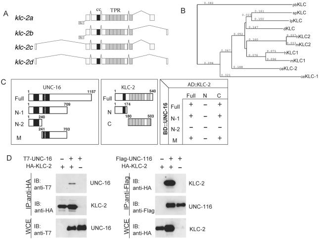
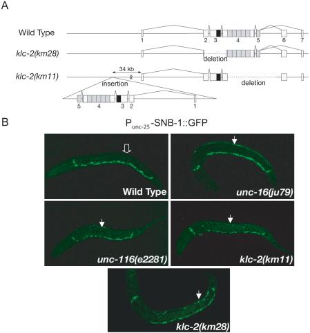
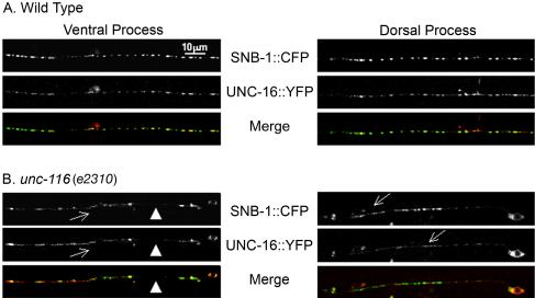
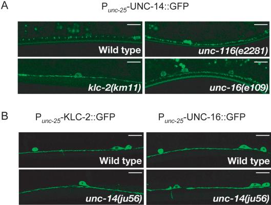
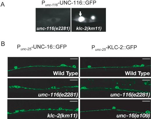
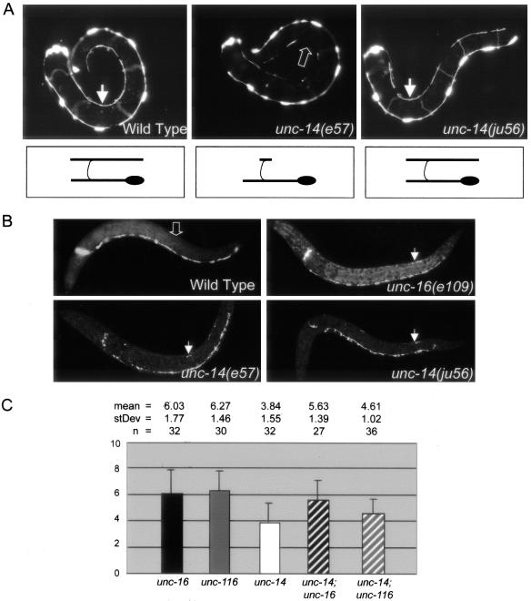
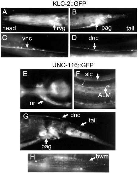
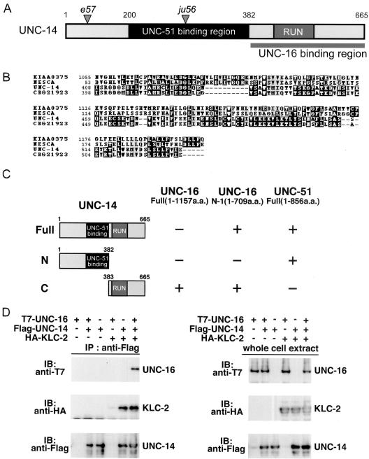
Kinesin-1 is a heterotetramer composed of kinesin heavy chain (KHC) and kinesin light chain (KLC). The Caenorhabditis elegans genome has a single KHC, encoded by the unc-116 gene, and two KLCs, encoded by the klc-1 and klc-2 genes. We show here that UNC-116/KHC and KLC-2 form a complex orthologous to conventional kinesin-1. KLC-2 also binds UNC-16, the C. elegans JIP3/JSAP1 JNK-signaling scaffold protein, and the UNC-14 RUN domain protein. The localization of UNC-16 and UNC-14 depends on kinesin-1 (UNC-116 and KLC-2). Furthermore, mutations in unc-16, klc-2, unc-116, and unc-14 all alter the localization of cargos containing synaptic vesicle markers. Double mutant analysis is consistent with these four genes functioning in the same pathway. Our data support a model whereby UNC-16 and UNC-14 function together as kinesin-1 cargos and regulators for the transport or localization of synaptic vesicle components.


Figures

Figure 1.
Figure 1.
KLC-2 binds UNC-16 and the UNC-116 KHC. (A) Structures of klc-2 isoforms. The deduced four alternative splicing forms of klc-2 are shown. The black and gray boxes indicate the regions encoding the coiled-coil domain and TPR motifs, respectively. The trans-splicing sites are also indicated. (B) Alignment dendrogram generated using the neighbor-joining method within CLUSTALX. Numeric values indicate branch lengths in terms of percent divergence. pb, Plectonema boryanum (cyanobacterium); sp, Strongylocentrotus purpuratus (sea urchin); lp, Loligo pealii (squid); d, Drosophila; h, human; m, mouse; and ce, C. elegans. (C) Interaction between KLC-2 and UNC-16 in the yeast two-hybrid system. Various regions of UNC-16 fused with the LexA DNA-binding domain (BD) are shown on the left. The regions of KLC-2 fused with the GAL4 activation domain (AD) are shown in the middle. The plus (+) and minus (-) indicate positive and negative interactions between the various regions of UNC-16 and KLC-2 in the chart on the right. (D) Association of KLC-2 with UNC-16 and UNC-116. HEK 293 cells were transfected with control vector (-), HA-KLC-2, T7-UNC-16, and Flag-UNC-116 as indicated. Cell lysates were immunoprecipitated (IP) with anti-HA (left) and anti-Flag (right) antibodies. Immunoprecipitates were immunoblotted (IB) with anti-T7 (left panel) and anti-HA (right) antibodies. The amounts of immunoprecipitated HA-KLC-2 and Flag-UNC-116 were determined with anti-HA (left) and anti-Flag (right) antibodies, respectively. Whole-cell extracts (WCE) were immunoblotted with anti-T7 (left) and anti-HA (right) antibodies to determine total amounts of T7-UNC-16 and HA-KLC-2, respectively.
Figure 2.
Figure 2.
klc-2 loss-of-function mutation affects synaptic vesicle marker localization. (A) Structure of the klc-2 gene. Exons are indicated by boxes with numbers. The black and gray boxes indicate regions encoding the coiled-coil domain and TPR motifs, respectively. The klc-2(km28) mutation is a 0.6 kb deletion. The klc-2(km11) mutant has two copies of the klc-2 gene. One deletes the entire regions encoding the TPRs, and the other is an insertion of a second copy of the klc-2 gene with a deletion removing the two alternative last exons. (B) SNB-1::GFP localization. SNB-1::GFP is localized only along the ventral DD processes in wild-type L1 larvae (open arrow in top left panel indicates lack of SNB-1::GFP along dorsal side), but along both the ventral and dorsal DD processes in unc-16(ju79), unc-116(e2281), klc-2(km28), and klc-2(km11) mutant L1 larvae (closed arrows indicate dorsal SNB-1::GFP in top right and bottom panels). All animals are positioned with anterior-left, posterior-right, ventral-down, and dorsal-up.
Figure 5.
Figure 5.
UNC-16 and SNB-1 localization is dependent on UNC-116. P unc-25-UNC-16::YFP and P unc-25-SNB-1::CFP (juEx832) were expressed in wild-type (A) and unc-116 mutant (B) animals. Shown are single plane scan confocal images, focusing on SNB-1::CFP. (A) UNC-16 is associated with SNB-1 puncta on the ventral and dorsal processes of wild-type animals (97.2 ± 0.9%). (B) In unc-116 (e2310) mutants, both UNC-16 and SNB-1 localization is altered. UNC-16 and SNB-1 are seen largely irregular and diffuse along the ventral process (arrows) and are often absent (arrowheads). In the rare case where SNB-1 expression was punctate, the incidence of colocalization with UNC-16 was decreased.
Figure 7.
Figure 7.
Localization of UNC-14 depends on kinesin-1. (A) Localization of UNC-14. P unc-25-UNC-14::GFP (kmEx821) was expressed in D-type neurons of wild-type, unc-116(e2281), klc-2(km11), and unc-16(e109) animals. Panels show localization of UNC-14::GFP in the ventral nerve processes of D-type neurons. UNC-14::GFP showed a punctate pattern in wild-type animals (top left). In unc-116(e2281), klc-2(km11), and unc-16(e109) mutant animals, UNC-14::GFP was diffused and irregular along the ventral nerve processes (top right and bottom panels). (B) Localization of KLC-2 and UNC-16 in unc-14 mutants. Punc-25-KLC-2::GFP (kmEx811) and Punc-25-UNC-16::GFP (kmEx823) were expressed in D-type neurons of wild-type and unc-14(ju56) mutant animals. Panels show localization of KLC-2::GFP (left panels) and UNC-16::GFP (right panels) in the ventral nerve processes of D-type neurons. Both KLC-2::GFP and UNC-16::GFP were evenly distributed in wild-type and unc-14(ju56) mutant animals. Scale, 10 μm
Figure 4.
Figure 4.
Localization dependence of UNC-16 and kinesin-1. (A) Subcellular localization of UNC-116::GFP. Panels show localization of Punc-116-UNC-116::GFP (juEx637) in an unc-116(e2281) rescued animal (left) and in a klc-2(km11) mutant animal (right). Arrow in right panel indicates perinuclear accumulation of UNC-116::GFP in the klc-2(km11) mutant cell. (B) Localization of UNC-16::GFP and KLC-2::GFP in kinesin-1 and unc-16 mutants. Punc-25-UNC-16::GFP (kmEx823) and Punc-25-KLC-2::GFP (kmEx811) were expressed in D-type neurons of wild-type, unc-116(e2281), klc-2(km11), and unc-16(e109) animals. Panels show localization of UNC-16::GFP (left panels) and KLC-2::GFP (right panels) in the ventral nerve processes of the D-type neurons. UNC-16::GFP is evenly distributed in wild-type animals (top left). In unc-116(e2281) and klc-2(km11) mutants, UNC-16::GFP is aggregated along the ventral nerve processes (middle and bottom left). KLC-2::GFP is evenly distributed in wild-type (top right) and in unc-16(e109) mutant animals (bottom right), but not uniform in unc-116(e2281) mutants (middle right). Scale, 10 μm.
Figure 8.
Figure 8.
unc-14 regulates synaptic vesicle localization and functions in the same pathway as unc-16 and unc-116. (A) DD cell morphology. Panels show L1 stage animals expressing Punc-25-GFP (juIs76) to visualize the morphology of the DD motor neurons. In wild-type animals, six DD cell bodies reside along the ventral nerve cord. Each extends a process along the ventral nerve cord, around the side of the body (commissures out of the focal plane), and along the dorsal nerve cord. The arrows in the left and right panels indicate fully extended dorsal DD processes in wild-type and unc-14(ju56) L1 animals. The open arrow in the middle panel indicates missing dorsal DD processes in an unc-14(e57) L1 animal. Drawings below the animals demonstrate the morphology of an individual DD motor neuron representative of each genotype. (B) SNB-1::GFP (juIs1) localization in L1 animals. Although wild-type L1s have no SNB-1::GFP along their dorsal DD processes (open arrow in top left panel), SNB-1::GFP is misaccumulated along the dorsal DD processes of unc-16 and unc-14 mutant L1 animals (closed arrows in top right and bottom panels). (C) Quantification of L1 dorsal SNB-1::GFP phenotype. The extent of dorsal SNB-1::GFP was scored in L1 animals on a scale from 0 to 25 (see Materials and Methods) in unc-16(e109), unc-116(e2281), and unc-14(ju56) single mutants, and in unc-14(ju56); unc-16(e109) and unc-14(ju56); unc-116(e2281) double-mutant animals. The mean, SD, and n are indicated above the graph. By Student's t-tests (2 tail/unpaired/unequal variance), the L1 SNB-1::GFP phenotypes of unc-16(e109) single mutants and unc-16(e109); unc-14(ju56) double mutants are not significantly different (p = 0.333), and the L1 SNB-1::GFP phenotypes of unc-14(ju56) single mutants and unc-14(ju56); unc-116(e2281) double mutants are not significantly different (p = 0.21). Although other pair combinations have significantly different mean scores, all differences are less than additive. p-values for other pair-wise comparisons are as follows: unc-16(e109) and unc-14(ju56) (p = 1.9e-6), unc-14(ju56) and unc-14(ju56); unc-16(e109) (p = 1.9e-5), unc-116(e2281) and unc-14(ju56) (p = 3.3e-8), and unc-116(e2281) and unc-14(ju56); unc-116(e2281) (p = 3.2e-6).
Figure 3.
Figure 3.
Expression of KLC-2 and UNC-116. (A-D) Expression pattern and localization of KLC-2::GFP. Panels show wild-type young adult animals expressing Pklc-2-KLC-2::GFP (kmEx801). Arrows indicate rvg, retrovesicular ganglion (A); pag, preanal ganglion (B); vnc, ventral nerve cord (C); and dnc, dorsal nerve cord (D). (E-H) Expression pattern and localization of UNC-116::GFP. Panels show unc-116(e2281) animals rescued by expression of Punc-116 UNC-116::GFP (juEx637). Arrows indicate nrn, nerve ring neuropile (E); ALM mechanosensory neuron and slnc, sublateral nerve cord (F); various cell types in the tail (G); and bwm, body wall muscle (H).
Figure 6.
Figure 6.
UNC-16 binds to the RUN domain region of UNC-14. (A) UNC-14 protein domain structure. The RUN domain and the UNC-16 binding region are shown; the shaded region shows the region that was previously reported to bind UNC-51 (Ogura et al., 1997). Positions of the e57 and ju56 early stop mutations are also indicated. (B) RUN domain sequence alignment. Multiple sequence alignment with human NESCA and conceptual translation of KIAA0375, C. elegans UNC-14, and C. briggsae CBG21923 RUN domains constructed with CLUSTALW and displayed by BOXSHADE. (C) Interaction of UNC-14 with UNC-16 and UNC-51 in the yeast two-hybrid system. UNC-16 N-1 and UNC-51 were constructed with the LexA DNA-binding domain (BD). Various regions of UNC-14 constructed with the GAL4 activation domain (AD) are shown on the left. Plus (+) and minus (-) indicate positive and negative interactions, respectively. (D) Coimmunopreciptation of UNC-16 and KLC-2 with UNC-14. HEK 293 cells were transfected with control vector (-), T7-UNC-16, Flag-UNC-14, and HA-KLC-2, as indicated. Cell lysates were immunoprecipitated (IP) with anti-Flag antibody (left panels). Immunoprecipitates were immunoblotted (IB) with anti-T7 (top panel) and anti-HA (middle panel) antibodies. The amounts of immunoprecipitated Flag-UNC-14 were determined with anti-Flag antibody (bottom panel). Whole-cell extracts were immunoblotted with anti-T7, anti-HA, and anti-Flag antibodies to determine total amounts of T7-UNC-16, HA-KLC-2, and Flag-UNC-14, respectively (right panels).

Similar articles

Cited by

References

    1. Akechi, M., Ito, M., Uemura, K., Takamatsu, N., Yamashita, S., Uchiyama, K., Yoshioka, K., and Shiba, T. (2001). Expression of JNK cascade scaffold protein JSAP1 in the mouse nervous system. Neurosci. Res. 39, 391-400. - PubMed
    1. Bowman, A. B., Kamal, A., Ritchings, B. W., Philp, A. V., McGrail, M., Gindhart, J. G., and Goldstein, L.S. (2000). Kinesin-dependent axonal transport is mediated by the sunday driver (SYD) protein. Cell 103, 583-594. - PubMed
    1. Brady, S. T. (1985). A novel brain ATPase with properties expected for the fast axonal transport motor. Nature 317, 73-75. - PubMed
    1. Brenner, S. (1974). The genetics of Caenorhabditis elegans. Genetics 77, 71-94. - PMC - PubMed
    1. Byrd, D. T., Kawasaki, M., Walcoff, M., Hisamoto, N., Matsumoto, K., and Jin, Y. (2001). UNC-16, a JNK-signaling scaffold protein, regulates vesicle transport in C. elegans. Neuron 32, 787-800. - PubMed

Publication types

MeSH terms

LinkOut - more resources