. 2005 Feb;16(2):446-57.
doi: 10.1091/mbc.e04-09-0842. Epub 2004 Nov 24.

A sorting nexin PpAtg24 regulates vacuolar membrane dynamics during pexophagy via binding to phosphatidylinositol-3-phosphate

Affiliations

A sorting nexin PpAtg24 regulates vacuolar membrane dynamics during pexophagy via binding to phosphatidylinositol-3-phosphate

Yoshitaka Ano et al. Mol Biol Cell. 2005 Feb.

Abstract

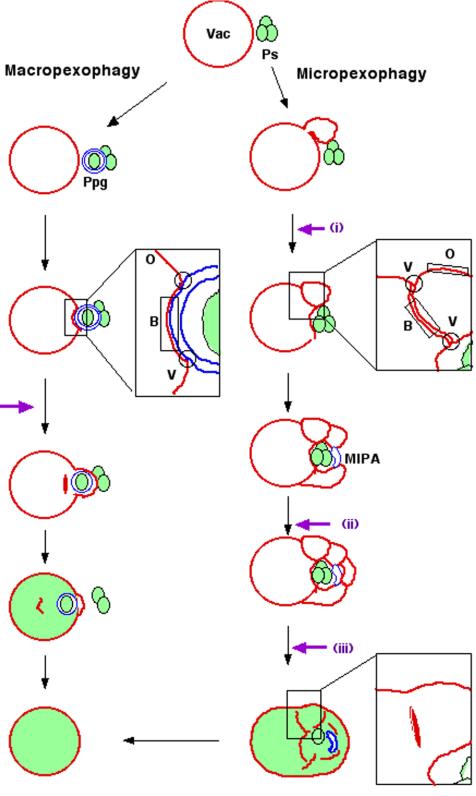
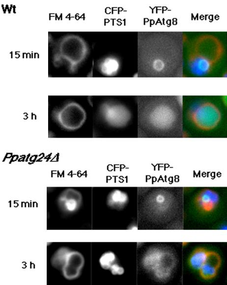
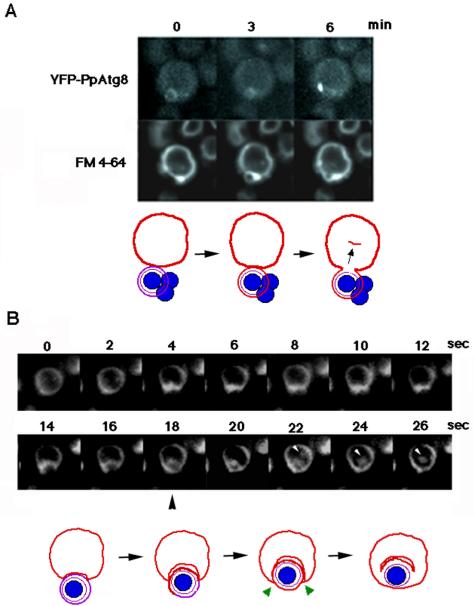
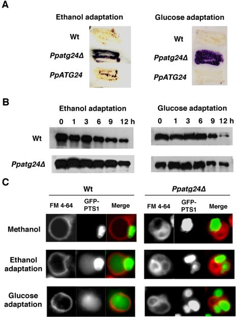
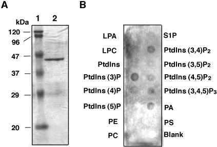
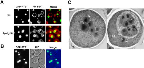
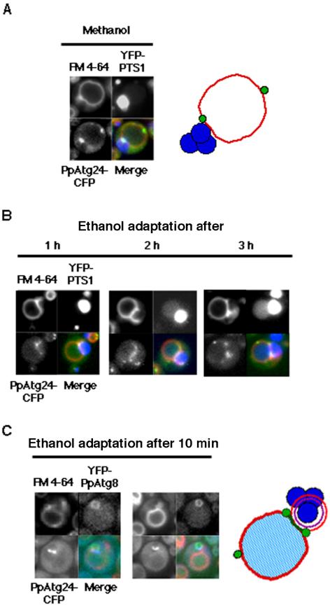
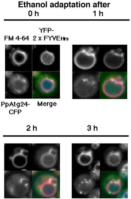
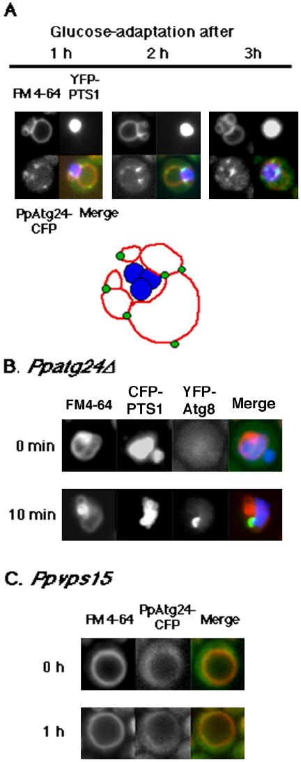
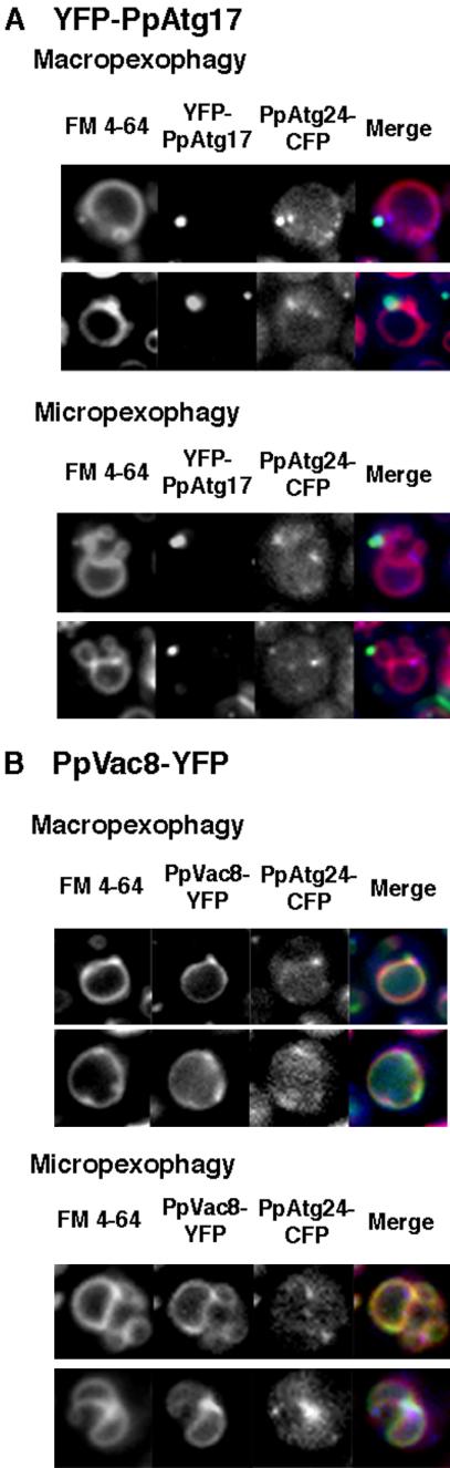
Diverse cellular processes such as autophagic protein degradation require phosphoinositide signaling in eukaryotic cells. In the methylotrophic yeast Pichia pastoris, peroxisomes can be selectively degraded via two types of pexophagic pathways, macropexophagy and micropexophagy. Both involve membrane fusion events at the vacuolar surface that are characterized by internalization of the boundary domain of the fusion complex, indicating that fusion occurs at the vertex. Here, we show that PpAtg24, a molecule with a phosphatidylinositol 3-phosphate-binding module (PX domain) that is indispensable for pexophagy, functions in membrane fusion at the vacuolar surface. CFP-tagged PpAtg24 localized to the vertex and boundary region of the pexophagosome-vacuole fusion complex during macropexophagy. Depletion of PpAtg24 resulted in the blockage of macropexophagy after pexophagosome formation and before the fusion stage. These and other results suggest that PpAtg24 is involved in the spatiotemporal regulation of membrane fusion at the vacuolar surface during pexophagy via binding to phosphatidylinositol 3-phosphate, rather than the previously suggested function in formation of the pexophagosome.


Figures

Figure 1.
Figure 1.
Schematic model for vacuolar membrane dynamics of two distinct pexophagic pathways and subdomains on the vacuolar membrane. Violet arrows represent possible fusion events occurring at the vacuolar membrane surface. Ps, peroxisome; Vac, vacuole; MIPA, micropexophagic apparatus; Ppg, pexophagosome; V, vertex domain; B, boundary domain; O, outside edge domain. Left: macropexophagy. A newly synthesized pexophagosome envelops a single peroxisome within a cluster, and subsequently its outer membrane fuses with vacuolar membrane. This fusion event could occur in two different ways: fusion at a contact point or fusion at a vertex. This figure represents the fusion at a vertex. Although fusion at the vertex involves internalization of the boundary region (as described in this figure), fusion at the contact point does not. Right: micropexophagy. Membrane fusion (or scission) at the vacuolar surface should occur at three different steps (indicated by violet arrows): i) septation of the vacuole; ii) completion of peroxisome engulfment; and iii) degradation of the septum.
Figure 6.
Figure 6.
Dynamics of YFP-PpAtg8 localization during macropexophagy in the wild-type and the Ppatg24Δ strain. After ethanol-adaptation for 15 min, both the wild-type (strain YAP0004) and the Ppatg24Δ (YAP2415) strains showed a ring structure representing a pexophagosome. After 3 h, while YFP-PpAtg8 fluorescence could be observed in the vacuolar lumen in wild-type cells, YFP fluorescence in the vacuolar lumen was not observed in the Ppatg24Δ strain.
Figure 2.
Figure 2.
Single-cell observation of FM 4-64-stained vacuolar membrane dynamics during macropexophagy. (A) Sequential images of YFP-PpAtg8 dynamics taken at 3-min intervals. (B) Sequential images taken at 2-s intervals. White arrowheads represent the internalized membrane showing Brownian movement within the vacuolar lumen. FM 4-64 diffuses into the pexophaogome membrane after the fusion (black arrowhead). Bottom panels: a scheme for vacuolar membrane dynamics deduced from the top panels. Red line, vacuolar membrane originally stained with FM 4-64; violet line, pexophagosome membrane; blue circle, peroxisome; green arrow heads, fusion points at the vertex.
Figure 3.
Figure 3.
PpATG24 disruption leads to deficient degradation of the peroxisomal enzyme Aox. (A) Remaining Aox activity after pexophagy induction. Wild-type (Wt; strain PPY12), Ppatg24Δ (strain YAP2401), and PpATG24 (strain YAP2403) cells grown on a methanol plate were adapted to glucose or ethanol for 12 or 48 h, respectively. Purple represents the persistence of the peroxisomal protein Aox, as detected by its activity. In wild-type cells (Wt), Aox was degraded after the ethanol adaptation (macropexophagy) and glucose adaptation (micropexophagy), whereas both pathways were impaired in the Ppatg24Δ mutant. By introduction of the PpATG24 gene into Ppatg24Δ, peroxisome degradation was restored completely (PpATG24). (B) Aox degradation during ethanol or glucose adaptation. The methanol-grown wild-type and Ppatg24Δ cells were transferred to glucose or ethanol medium and harvested after the indicated times. Cell lysates were then subjected to immunoblot analysis using anti-Aox antibody. A decrease in the intensity of the Aox signal was retarded in the Ppatg24Δ strain. The decrease of signal in Ppatg24Δ strain was due to the dilution of Aox-protein by cell growth after the medium shift. (C) Fluorescent images of wild-type (STWI) and Ppatg24Δ (YAP2402) cells labeled with GFP-PTS1 and FM 4-64. Pexophagy was detected by diffusion of GFP-PTS1 in the vacuolar lumen in the wild-type cells after ethanol- or glucose-adaptation for 3 h. Diffusion of GFP-PTS1 in the vacuolar lumen was not observed in Ppatg24Δ strain.
Figure 4.
Figure 4.
Specific binding of PpAtg24 phox homology (PX) domain to phosphatidylinositol-3-phosphate (PtdIns(3)P). (A) Purity of the GST-PpAtg24 PX domain detected on SDS-PAGE. The GST-fused PpAtg24 PX domain was purified using an E. coli Rosetta DE3 by GSTrap FF column. The purity of the purified protein was determined by Coomassie staining. Lane 1, molecular weight markers; lane 2, purified GST-PpAtg24 PX domain protein. (B) Protein-lipid overlay assay. Purified GST-PpAtg24 PX domain was subjected to PIP-Strip using anti-GST antiserum for immunoblot analysis. LPA, lysophosphatidic acid; LPC, lysophosphocholine; PtdIns, phsophatidylinositol; PE, phosphatidylethanolamine; PC, phsophatidylcholine; S1P, sphingosine-1-phosphate; PA, phosphatidic acid; PS, phosphatidylserine.
Figure 5.
Figure 5.
The deletion of PpATG24 causes aberrant vacuole morphology and peroxisome cluster impairment in methanol-grown cells. (A) Methanol-grown wild-type (STWI) and Ppatg24Δ (YAP2402) cells were labeled with GFP-PTS1 and FM 4-64 and were observed by fluorescence microscopy. Wild-type cells formed a large, round vacuole before pexophagy. In contrast, Ppatg24Δ cells showed aberrant vacuole morphology. Wild-type methanol-grown cells always contained one peroxisomal cluster. (B) Peroxisomal clustering was inhibited in some Ppatg24Δ cells (strain YAP2402). (C) Two electron micrographs of methanol-grown Ppatg24Δ cells. Before induction of pexophagy, cells showed extensive engulfment of peroxisomes and cytosol by vacuolar membranes. Note that the mitochondrion was not wrapped in the vacuolar membrane, and one peroxisome was isolated from the other clusters (indicated by an arrowhead). P, peroxisome; V, vacuole; M, mitochondrion; N, nucleus.
Figure 7.
Figure 7.
Intracellular behavior of PpAtg24 during macropexophagy. Pichia cells expressing PpAtg24-CFP and YFP-PTS1 in Ppatg24Δ (YAP2405) were transferred from methanol medium to ethanol medium for induction of macropexophagy. (A and B) Superimposed images are shown with (red) FM4-64, (blue) YFP-PTS1, and (green) PpAtg24-CFP signal. (A) Methanol-grown cell. Left, fluorescent images; right, representative localization of PpAtg24-CFP. (B) Fluorescent images after ethanol-adaptation. A major portion of the PpAtg24-CFP spot fluorescence was juxtaposed to the peroxisome cluster and pexophagosome, at the vertex and boundary region. Fusion could be detected by the flow of YFP-PTS1 into the vacuolar lumen after 2 h. (C) PpAtg24-CFP-expressing cells were costained with YFP-PpAtg8 (strain YAP2411) and FM 4-64 (left). In these cells, the fusion was not detected because we could observe a ring-shaped PpAtg8 fluorescence (see Figure 2A). Representative localization of PpAtg24-CFP during macropexophagy (right). Green, PpAtg24-CFP; red, vacuole membrane; blue, peroxisomes.
Figure 8.
Figure 8.
Localization of PtdIns(3)P and PpAtg24 during macropexophagy. Methanol-grown Pichia cells (YAP2406) expressing the YFP-2xFYVE domain from mouse Hrs (PtdIns(3)P marker) and PpAtg24-CFP were transferred from methanol medium to ethanol medium for induction of macropexophagy. Merged images are stained with (red) FM4-64, (green) YFP-2xFYVEHrs domain, and (blue) PpAtg24-CFP.
Figure 9.
Figure 9.
Intracellular behavior of PpAtg24 during micropexophagy. (A) Top: Ppatg24Δ (YAP2405) Pichia cells expressing PpAtg24-CFP and YFP-PTS1, YFP-tagged peroxisomal targeting signal were transferred from methanol medium to glucose medium for induction of micropexophagy. Superimposed images showing (red) FM4-64, (blue) YFP-PTS1, and (green) PpAtg24-CFP signal. A major part of the PpAtg24-CFP spot fluorescence was localized to the vertex region or tips of the septating vacuole. Bottom: representative localization of PpAtg24-CFP during micropexophagy. (B) The extended cup-like YFP-PpAtg8 fluorescence appeared after 10 min of micropexphagy induction, indicating that the MIPA-like structure was formed in Ppatg24Δ cells. (C) Localization of PpAtg24-CFP in the Ppvps15 mutant strain. Wild-type (YAP2404) and PpAtg24-CFP-expressing Ppvps15 (YAP2410) cells were shifted to glucose medium for 60 min. The vacuole failed to invaginate or septate in the vps15 mutant.
Figure 10.
Figure 10.
A portion of PpAtg24-CFP spot fluorescence colocalized with YFP-Atg17 and PpVac8-YFP during pexophagy. (A) YFP-PpAtg17 and (B) PpVac8-YFP were introduced into the PpAtg24-CFP-expressing strain, yielding the YAP2413 strain and the YAP2414 strain, respectively. Macropexophagy or micropexophagy was induced in these cells.

Similar articles

Cited by

References

    1. Ago, T., Takeya, R., Hiroaki, H., Kuribayashi, F., Ito, T., Kohda, D., and Sumimoto, H. (2001). The PX domain as a novel phosphoinositide-binding module. Biochem. Biophys. Res. Commun. 287, 733-738. - PubMed
    1. Ausubel, F. M., Brent, R., Kingston, R. E., Moore, D. D., Seidman, J. G., Smith, J. A., and Struhl, K. (eds.) (1987). Current Protocols in Molecular Biology, New York: Greene Publishing Associates and Wiley-Interscience.
    1. Baba, M., Osumi, M., Scott, S. V., Klionsky, D. J., and Ohsumi, Y. (1997). Two distinct pathways for targeting proteins from the cytoplasm to the vacuole/lysosome. J. Cell Biol. 139, 1687-1695. - PMC - PubMed
    1. Cheever, M. L., Sato, T. K., de Beer, T., Kutateladze, T. G., Emr, S. D., and Overduin, M. (2001). Phox domain interaction with PtdIns(3)P targets the Vam7 t-SNARE to vacuole membranes. Nat. Cell Biol. 3, 613-618. - PubMed
    1. Gillooly, D. J., Morrow, I. C., Lindsay, M., Gould, R., Bryant, N. J., Gaullier, J. M., Parton, R. G., and Stenmark, H. (2000). Localization of phosphatidylinositol 3-phosphate in yeast and mammalian cells. EMBO J. 19, 4577-4588. - PMC - PubMed

Publication types

MeSH terms

LinkOut - more resources