. 2004 Nov 25:5:46.
doi: 10.1186/1471-2474-5-46.

Nodular osteochondrogenic activity in soft tissue surrounding osteoma in neurogenic para osteo-arthropathy: morphological and immunohistochemical study

Affiliations

Nodular osteochondrogenic activity in soft tissue surrounding osteoma in neurogenic para osteo-arthropathy: morphological and immunohistochemical study

T Youssefian et al. BMC Musculoskelet Disord. .

Abstract

Background: Neurogenic Para-Osteo-Arthropathy (NPOA) occurs as a consequence of central nervous system injuries or some systemic conditions. They are characterized by bone formation around the main joints.

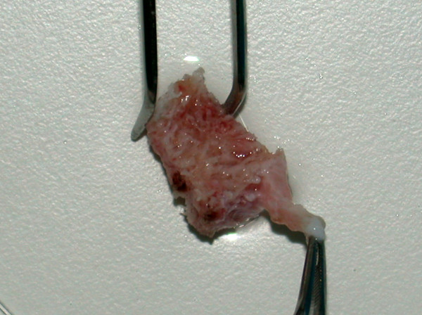
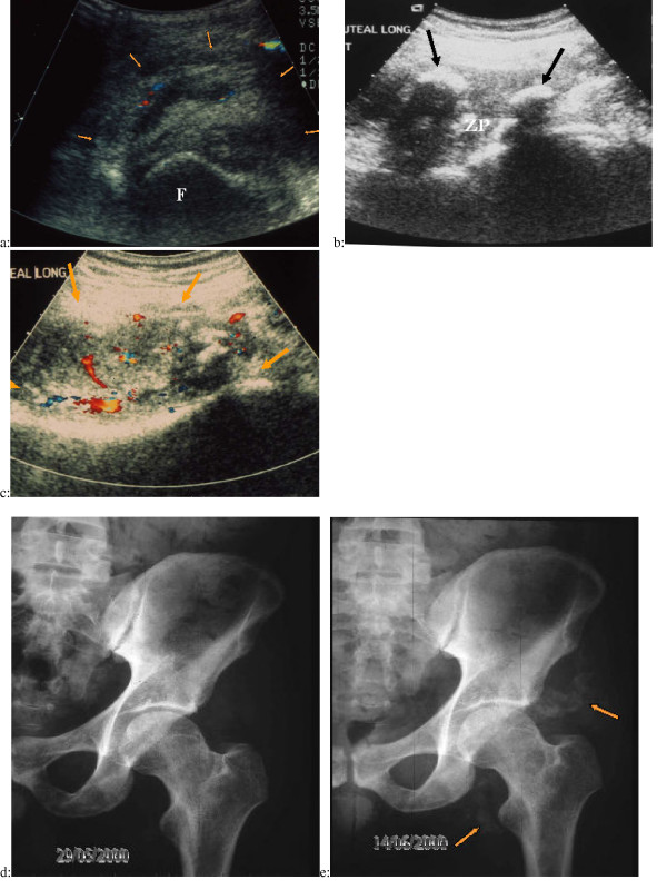
Methods: In order to define some biological features of NPOAs, histological and immunohistological studies of the soft tissue surrounding osteoma and Ultrasound examination (US) of NPOA before the appearance of abnormal ossification on plain radiographs were performed.

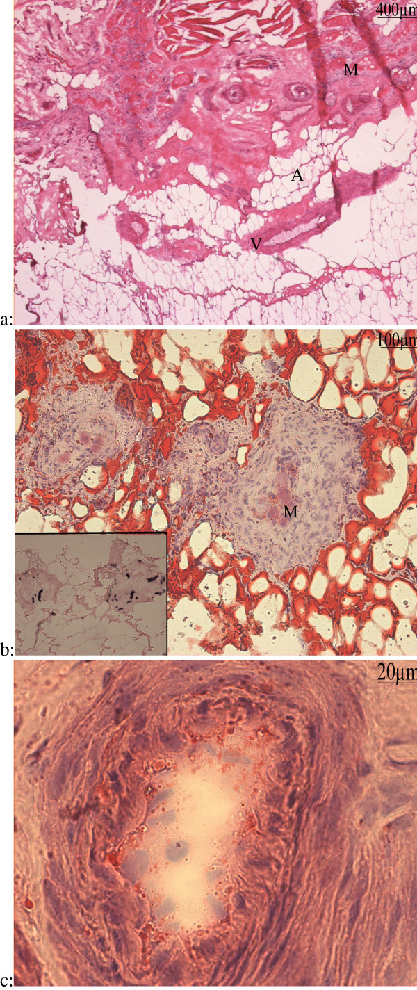
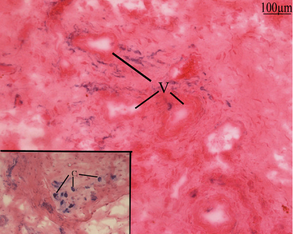
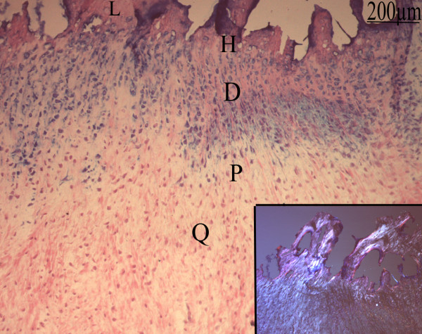
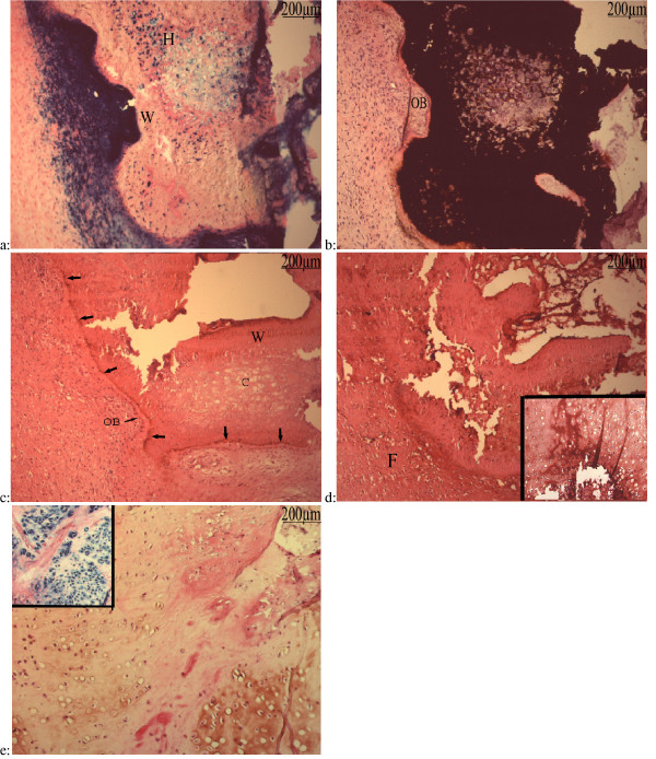
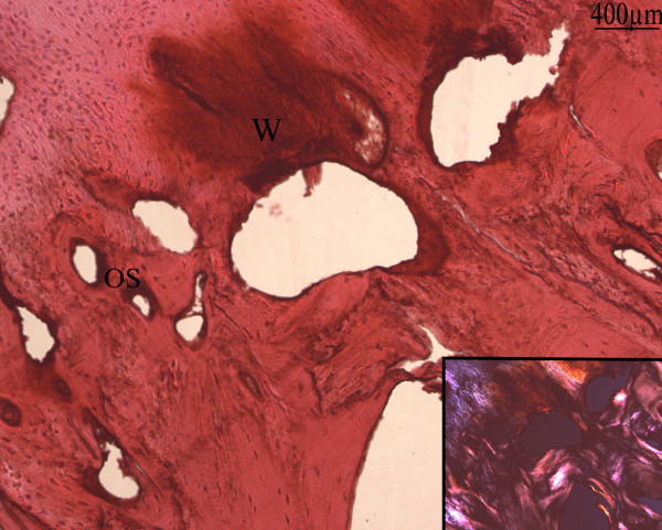
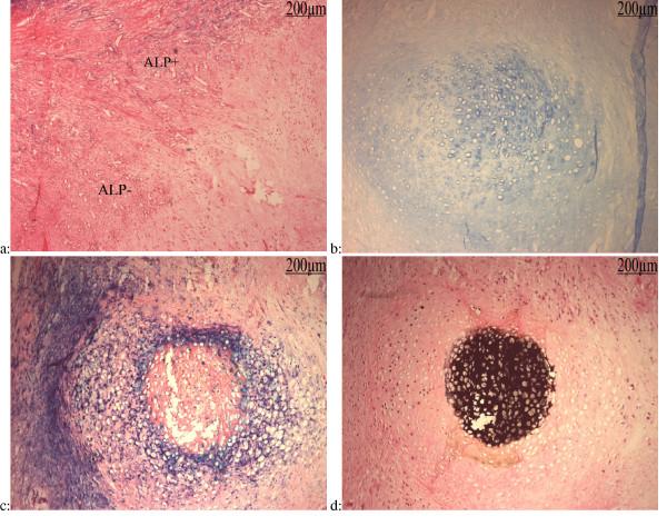
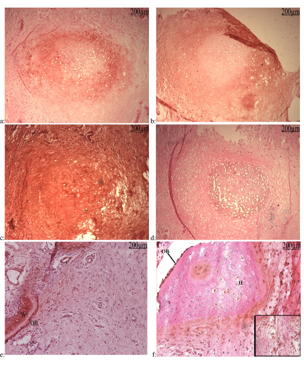
Results: We have observed a great number of ossifying areas scattered in soft tissues. US examination have also shown scattered ossifying areas at the early stage of ossification. A high osteogenic activity was detected in these tissues and all the stages of the endochondral process were observed. Mesenchymal cells undergo chondrocytic differentiation to further terminal maturation with hypertrophy, which sustains mineralization followed by endochondral ossification process.

Conclusion: We suggest that periosteoma soft tissue reflect early stage of osteoma formation and could be a model to study the mechanism of osteoma formation and we propose a mechanism of the NPOA formation in which sympathetic dystony and altered mechanical loading induce changes which could be responsible for the cascade of cellular events leading to cartilage and bone formation.


Figures

Figure 1
Figure 1
Dissection of part of the soft tissue surrounding a piece of osteoma.
Figure 2
Figure 2
a: Frozen section of soft tissue from a 72 months hip NPOA: Hematoxylin eosin (h&e): Thrombotic vessels (V), more or less advanced vacuolization/degeneration of muscular fibers (M), and adipous tissue (A). b: Oil Red-O staining : Degenerated muscle fibers (M) stained by eosin were embedded in a strongly hyperplastic perimysium. This structure was itself located inside an adipous tissue whose appearance and compartmentalization by endomysium-like sheets of cell layers suggests a muscular origin. Inset: ALP activity: the same area showed an intense ALP activity in some cells in hyperplastic perimysium. c: Hematoxylin eosin (h&e) staining showed hyperplastic intima and media.
Figure 3
Figure 3
Frozen sections of soft tissue of an 8 months hip NPOA: Blue Alkaline Phosphatase (ALP) activity counter stained with nuclear red-eosin: perivascular cells show high ALP activity near by vessels (V) and some of these seems to migrate from these areas. Inset: Cluster and isolated rounded cells (C) with high ALP showed a more advanced stage of differentiation.
Figure 4
Figure 4
Frozen section of soft tissue of an 8 months hip NPOA: Blue Alkaline Phosphatase (ALP) activity counter stained with nuclear red-eosin: The successive stage of chondrocytes differentiation were observed: quiescent (Q), proliferative (P), prehypertrophic (D), hypertrophic (H) and mineralization zones. Inset: Lamellar bone deposition (L) is visualized with polarized light.
Figure 5
Figure 5
Frozen sections of soft tissue of a 5 months elbow NPOA: a: Blue Alkaline Phosphatase (ALP) activity counter stained by nuclear red-eosin: A very strong ALP activity in multilayered cells surrounded a peripherically mineralized area. This area was made of woven bone (W) deposed on an hypertrophic cartilage (H) centered by prehypertrophic chondrocytes. Hypertrophic and prehypertrophic chondrocytes in the non-mineralized matrix displayed ALP activity. b: Vonkossa staining of a next section counter stained by h&e: Von Kossa stain was a negative image of the ALP activity. Osteoid matrix was deposed upon the calcified cartilage matrix and underwent mineralization. Palissade-arranged cells lined this osteoid and displayed morphological image of osteoblast-like cells (OB). c: Oscteocalcin immunolabelling of a next section of the same specimen counter stained by h&e: Osteocalcin immunoreactivity in the eosinophilic osteoid at the border of the mineralized woven bone and sligtly on osteoblast-like cells (OB) lining this zone. Slight labelling was also present in the woven bone (W), but not in the cartilage (C) areas. d: Type X collagen immunolabelling of a next section of the same specimen counter stained by h&e: Type X collagen immunoreactivity was found in the mineralized cartilage area up to the woven bone. It also streched over most of the fibrous tissue (F) surrounding the mineralized area. Inset: It was also present in hypertrophic chondrocytes and their matrix. e: Type II collagen immunolabelling of this specimen conter stain by h&e: The matrix of the columnar cartilage was immunolabelled with type II collagen antibody. Remnants of eosinophilic degenerated muscle fibers were interspersed among the cartilage. Inset: The type II colllagen immunoreactive areas displayed an heavy ALP activity.
Figure 6
Figure 6
Frozen section of soft tissue of a 24 months hip NPOA: Type I collagen immunolabelling counter stained by h&e: Type I collagen was the main constituant of the matrix in the primary osteons (OS) and non organized woven bone (W). Inset: lamellar bone deposition observed by polarized light
Figure 7
Figure 7
Serial sections of soft tissue of 24 months hip NPOA: a: ALP activity counter stained with nuclear red-eosin: Slide 118; Important angiogenesis encircles avascular areas. Many of these vessels express ALP activity (ALP+). b: Alcian bleu pH = 1 staining: Slide 70: chondroitin sulfate accumulation in cartilage. c: ALP activity counter stained with nuclear red-eosin: Slide 71. d: Von Kossa staining countre stained with h&e: Slide 65.
Figure 8
Figure 8
Immunological study of serial sections: a: Type II Collagen immunolabellingcountre stained with h&e: Slide 69: Type II collagen had the same pattern of ALP activity of nodule showing prehypertrophic chondrocytes in nonmineralized zone. b: Type I collagen Immunolabelling countre stained with h&e: Slide 72; Type I collagen expression encercled the mineralized nodule. c: Type X collagen immunolabelling countre stained with h&e:Slide 73: Type X collagen was expressed by most of cells and distributed in their matrix. d: OCN Immunolabelling countre stained with h&e: Slide 64: OCN was expressed by hypertrophic chondrocytes. e: OCN immunolabelling counter stained with h&e: Activated osteoblasts (OB) and woven bone (W) are strongly labelled. Some capillaries (C) near by these areas express osteocaline too. f: VEGF immunolabelling counter stained with h&e: VEGF was expressed by some hypertrophic chondrocytes (H). Activated and non activated osteoblasts-like cells (OB) lining the cartilage express also VEGF. Inset: Clustered and isolated cells in the matrix, surrounding hypertrophic chondrocytes, which could be destinated to capillary or osteoblast formation, are also labelled by VEGF.
Figure 9
Figure 9
Ultrasound and color Doppler examination and digital radiographs of suspected NPOA a: Axial US view combined with color Doppler of the anterior side of the left hip in a paraplegic patient presenting acute limitation and inflammation of this joint. The striation of the psoas iliaque muscle, normally detectable at the anterior part of the hip joint with US examination, has disappeared. A relatively well defined mass (orange arrows) is detectable at the anterior part of the left femoral head (F). This mass is very heterogeneous with mixed hypo and hyper echoic areas. Color Doppler enables visualization of vessels in the mass (red and blue Doppler signals). A mass effect is visible on the femoral vessels (top right of the view). b: Same patient, one week later, axial US view of the posterior side of the left hip. The classical zone phenomenon (ZP) is detectable with a central hypoechoic area surrounded by hyper echoic nodules with posterior attenuation(black arrows). c: Axial US examination at the same day combined with color Doppler view of the posterior side of the left hip. A posterior mass (orange arrows) is also visible in the gluteal muscles, very heterogeneous with mixed hypo and hyper echoic areas. Color Doppler reveals large vessels in the mass (red and blue Doppler signals). d: Plain radiographs of the left hip obtained the same day as first US examination: Any sign of ossification is visible while a well defined mass is detected by US examination. e: Plain radiographs of the left hip obtained two weeks after: Early anterior and posterior NPOA ossification is only slightly visible two weeks (Orange arrows) after the initial clinical signs whereas the US examination was initially positive.

Similar articles

Cited by

  • Macrophage-derived oncostatin M contributes to human and mouse neurogenic heterotopic ossifications.
    Torossian F, Guerton B, Anginot A, Alexander KA, Desterke C, Soave S, Tseng HW, Arouche N, Boutin L, Kulina I, Salga M, Jose B, Pettit AR, Clay D, Rochet N, Vlachos E, Genet G, Debaud C, Denormandie P, Genet F, Sims NA, Banzet S, Levesque JP, Lataillade JJ, Le Bousse-Kerdilès MC. Torossian F, et al. JCI Insight. 2017 Nov 2;2(21):e96034. doi: 10.1172/jci.insight.96034. JCI Insight. 2017. PMID: 29093266 Free PMC article.

References

    1. Chantraine A, Nusgens B, Lapiere CM. Biochemical analysis of heterotopic ossification in spinal cord injury patients. Paraplegia. 1995;33:398–401. - PubMed
    1. Ebinger T, Roesch M, Kiefer H, Kinzl L, Schulte M. Influence of etiology in heterotopic bone formation of the hip. J Trauma. 2000;48:1058–1062. doi: 10.1097/00005373-200006000-00010. - DOI - PubMed
    1. Dejerine A, Cellier A. Paraosteoarthropathies of paraplegic patients by spinal cord lesion. Clinical and roentgenographic study. Clin Orthop. 1991;263:3–12. - PubMed
    1. Richards AM, Klaassen MF. Heterotopic ossification after severe burns: a report of three cases and review of the literature. Burns. 1997;23:64–68. doi: 10.1016/S0305-4179(96)00074-5. - DOI - PubMed
    1. Taly AB, Nair KP, Kumar MV, Jayakumar PN, Vasudev MK, Ravishankar D, Kalaivant PL, Padankatty BS, Murali T. Heterotopic ossification in non-traumatic myelopathies. Spinal Cord. 1999;37:47–49. doi: 10.1038/sj.sc.3100751. - DOI - PubMed

MeSH terms