Review
. 2004 Dec 8;343(1):23-40.
doi: 10.1016/j.gene.2004.08.010.

Lineage-specific and ubiquitous biological roles of the mammalian transcription factor LSF

Affiliations
Review

Lineage-specific and ubiquitous biological roles of the mammalian transcription factor LSF

Jelena Veljkovic et al. Gene. .

Abstract

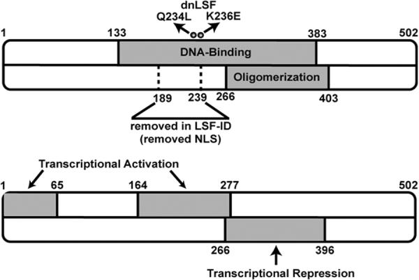
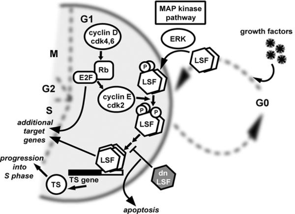
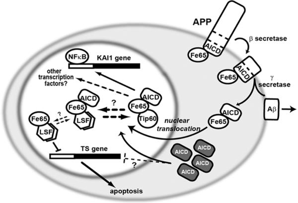
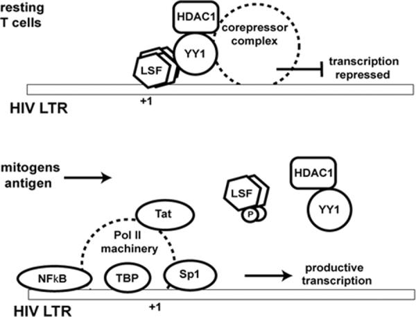
Transcriptional regulation in mammalian cells is driven by a complex interplay of multiple transcription factors that respond to signals from either external or internal stimuli. A single transcription factor can control expression of distinct sets of target genes, dependent on its state of post-translational modifications, interacting partner proteins, and the chromatin environment of the cellular genome. Furthermore, many transcription factors can act as either transcriptional repressors or activators, depending on promoter and cellular contexts [Alvarez, M., Rhodes, S.J., Bidwell, J.P., 2003. Context-dependent transcription: all politics is local. Gene 313, 43-57]. Even in this light, the versatility of LSF (Late SV40 Factor) is remarkable. A hallmark of LSF is its unusual DNA binding domain, as evidenced both by lack of homology to any other established DNA-binding domains and by its DNA recognition sequence. Although a dimer in solution, LSF requires additional multimerization with itself or partner proteins in order to interact with DNA. Transcriptionally, LSF can function as an activator or a repressor. It is a direct target of an increasing number of signal transduction pathways. Biologically, LSF plays roles in cell cycle progression and cell survival, as well as in cell lineage-specific functions, shown most strikingly to date in hematopoietic lineages. This review discusses how the unique aspects of LSF DNA-binding activity may make it particularly susceptible to regulation by signal transduction pathways and may relate to its distinct biological roles. We present current progress in elucidation of both tissue-specific and more universal cellular roles of LSF. Finally, we discuss suggestive data linking LSF to signaling by the amyloid precursor protein and to Alzheimer's disease, as well as to the regulation of latency of the human immunodeficiency virus (HIV).


Figures

Figure 1
Figure 1. Identified proteins in the LSF subfamily
Both human and mouse genomes contain three genes encoding LSF subfamily members. A splicing variant mRNA lacking one exon of the LSF gene encodes LSF-ID (LBP- 1d). A splicing variant mRNA containing an extra exon of the LBP-1a/b gene encodes the protein LBP-1b. While the LSF and LBP-1a/b genes are ubiquitously expressed, the third paralog, LBP9, exhibits a more limited expression profile; LBP9 also apparently lacks the transcriptional activation region conserved between LSF and LBP-1a/b. All LSF subfamily members oligomerize with each other.
Figure 2
Figure 2. DNA recognition sequences for LSF and GRH subfamily members
Known binding sites for mammalian LSF (Frith et al., 2001) (panel A) and D. melanogaster and C. elegans GRH (Venkatesan et al., 2003) (panel B) proteins were compiled. The resulting count matrices are presented as pictograms, presented according to the guidelines at http://www.genes.mit.edu/pictogram.html. The LSF and GRH pictograms are aligned in order to emphasize the similarity in sequence, with the LSF binding site containing two direct repeats of the sequence.
Figure 3
Figure 3. Structural/functional regions of LSF
The regions containing LSF DNA-binding, oligomerization, transcriptional activation and transcriptional repression activities are indicated in gray. The two amino acid substitutions within the DNA binding domain (Q234L and K236E) comprise the dominant negative mutant (dnLSF) and render the protein incapable of binding DNA while still being able to oligomerize. The deletion in LSF-ID is also shown, which causes the protein to be localized to the cytoplasm; therefore aa 189–239 contribute to nuclear localization.
Figure 4
Figure 4. Role of LSF in cell cycle progression via activation of the thymidylate synthase (TS) gene
Upon entry into G1, cyclin D/cdk4 and cyclin D/cdk6 complexes phosphorylate the retinoblastoma protein (Rb), allowing release of transcription factor E2F family members. A major role for E2F in cell cycle progression is to upregulate cyclin E/cdk2. In a parallel pathway, upon stimulation of cells in the resting (G0) state with growth factors, LSF is immediately and quantitatively phosphorylated by Erk, a central kinase in the mitogen activated protein kinase (MAP kinase) pathway. LSF is subsequently phosphorylated by cdk2, as well. One major target gene of active LSF is the thymidylate synthase (TS) gene. TS is essential for DNA replication in S phase. Expression of dnLSF prevents binding of LSF to target sites, and the lack of TS causes cells to undergo apoptosis. E2F, and presumably LSF, target additional genes for regulation during cell cycle progression into S phase. If growth factors are removed from the cellular environment, cells exit the cell cycle in G1, returning to the G0 state.
Figure 5
Figure 5. Potential role for LSF in mediating signal transduction from APP in the nucleus
The intracellular domain of the transmembrane protein APP (amyloid precursor protein) interacts with a number of signaling proteins, including Fe65. APP cleavages produce AICD (APP intracellular C-terminal domain) and Aβ (β amyloid) peptide. Aβ is released extracellularly and is responsible for plaque formation in Alzheimer's disease. AICD is released intracellularly, where Fe65 facilitates its translocation into the nucleus. Nuclear Fe65-AICD interacts with Tip60 and LSF, perhaps in competition with each other, as indicated. Fe65 alone inhibits LSF transactivation on the TS promoter, although whether the ternary complex with AICD is also inhibitory is not known. One target of the Fe65-AICD-Tip60 complex is the tetraspanin KAI1 gene, although many other target genes are likely, including through factors other than NF-κB. Overexpression of AICD (dark boxes) increased neuronal apoptosis, presumably in part by increased nuclear translocation and misregulation of target genes, perhaps including TS.
Figure 6
Figure 6. LSF represses transcription from the HIV LTR: Model of a role for LSF in viral latency
In resting T cells, transcription from the HIV LTR may be repressed by a complex including LSF, YY1 and histone deacetylase 1 (HDAC1). Upon stimulation of T cells by mitogens or by a specific antigen, the LSF-containing complex would be released, permitting assembly of the activating transcription factors, including Sp1, NF-κB, TATA-binding protein (TBP), and the rest of the RNA pol II machinery. Displacement of the repressive complex may be triggered by phosphorylation of LSF. Upon induction of HIV gene expression, Tat is produced, which further enhances productive transcription of the HIV genome.

References

    1. Algar E. A review of the Wilms' tumor 1 gene (WT1) and its role in hematopoiesis and leukemia. J Hematother Stem Cell Res. 2002;11:589–599. - PubMed
    1. Alvarez M, Rhodes SJ, Bidwell JP. Context-dependent transcription: all politics is local. Gene. 2003;313:43–57. - PubMed
    1. Attardi LD, Tjian R. Drosophila tissue-specific transcription factor NTF-1 contains a novel isoleucine-rich activation motif. Genes Dev. 1993;7:1341–1353. - PubMed
    1. Baek SH, Ohgi KA, Rose DW, Koo EH, Glass CK, Rosenfeld MG. Exchange of N-CoR corepressor and Tip60 coactivator complexes links gene expression by NF-κB and β-amyloid precursor protein. Cell. 2002;110:55–67. - PubMed
    1. Barnhart KM, Kim CG, Banerji SS, Sheffery M. Identification and characterization of multiple erythoid cell proteins that interact with the promoter of the murine α-globin gene. Mol Cell Biol. 1988;8:3215–3226. - PMC - PubMed

Publication types

LinkOut - more resources