

Review
doi: 10.1016/j.ccm.2004.06.003.
Cryptogenic organizing pneumonia
Affiliations
- PMID: 15564018
- PMCID: PMC7119066
- DOI: 10.1016/j.ccm.2004.06.003
Item in Clipboard
Review
Cryptogenic organizing pneumonia
Clin Chest Med.
2004 Dec.
Abstract
Cryptogenic organizing pneumonia is a rare, distinct disorder that is sufficiently different from the other diseases in the group of idiopathic interstitial pneumonias to be designated as a separate entity. In its most typical presentation, it is characterized by dyspnea and cough, with multiple patchy alveolar opacities on pulmonary imaging. Definite diagnosis is obtained by the finding of buds of granulation tissue in the distal airspaces at lung biopsy. No cause (as infection, drug reaction, or associated disease as connective tissue disease) is found. Corticosteroid treatment is rapidly effective, but relapses are common on reducing or stopping treatment.
Figures
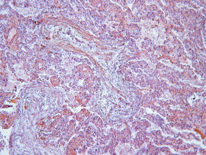
Buds of granulation tissue in the lumen of the distal airspaces.
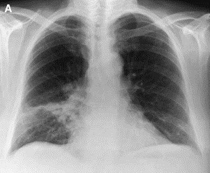
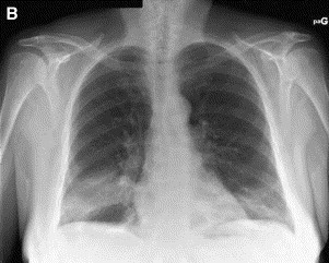
Chest radiograph in a patient with typical COP. (A) Patchy alveolar opacity of right lower lobe. (B) Six days later, the distribution of the right lower lobe opacity changed, and a new contralateral basal opacity appeared.
Chest radiograph in a patient with typical COP. (A) Patchy alveolar opacity of right lower lobe. (B) Six days later, the distribution of the right lower lobe opacity changed, and a new contralateral basal opacity appeared.
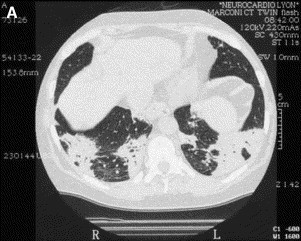
HRCT in typical COP. (A) Patchy bilateral opacities in the lower lobes. (B) Bilateral extensive consolidation with air bronchogram in the lower lobes.

HRCT in typical COP. (A) Patchy bilateral opacities in the lower lobes. (B) Bilateral extensive consolidation with air bronchogram in the lower lobes.
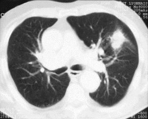
HRCT in solitary focal COP shows a pseudo-neoplastic mass.
References
-
- American Thoracic Society/European Respiratory Society Classification of the idiopathic interstitial pneumonias: international multidisciplinary consensus. American Thoracic Society/European Respiratory Society. Am J Respir Crit Care Med. 2002;165:277–304. - PubMed
-
- Colby T.V. Pathologic aspects of bronchiolitis obliterans organizing pneumonia. Chest. 1992;102:38S–43S. - PubMed
-
- Kitaichi M. Differential diagnosis of bronchiolitis obliterans organizing pneumonia. Chest. 1992;102:44S–49S. - PubMed
-
- Uner A.H., Rozum-Slota B., Katzenstein A.L. Bronchiolitis obliterans-organizing pneumonia (BOOP)-like variant of Wegener's granulomatosis: a clinicopathologic study of 16 cases. Am J Surg Pathol. 1996;20:794–801. - PubMed
-
- Perez de Llano L.A., Racamonde A.V., Bande M.J., Piquer M.O., Nieves F.B., Feijoo A.R. Bronchiolitis obliterans with organizing pneumonia associated with acute Coxiella burnetii infection. Respiration (Herrlisheim) 2001;68:425–427. - PubMed
Publication types
MeSH terms
LinkOut - more resources
Full Text Sources
