. 2004 Dec;78(24):13489-500.
doi: 10.1128/JVI.78.24.13489-13500.2004.

Two different molecular defects in the Tva receptor gene explain the resistance of two tvar lines of chickens to infection by subgroup A avian sarcoma and leukosis viruses

Affiliations

Two different molecular defects in the Tva receptor gene explain the resistance of two tvar lines of chickens to infection by subgroup A avian sarcoma and leukosis viruses

Daniel Elleder et al. J Virol. 2004 Dec.

Abstract

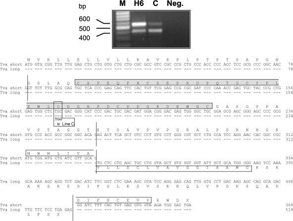
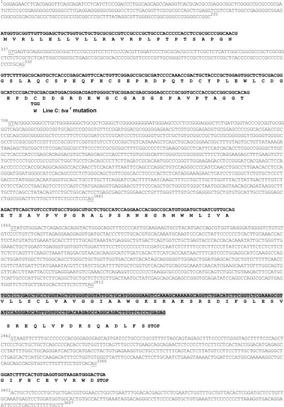
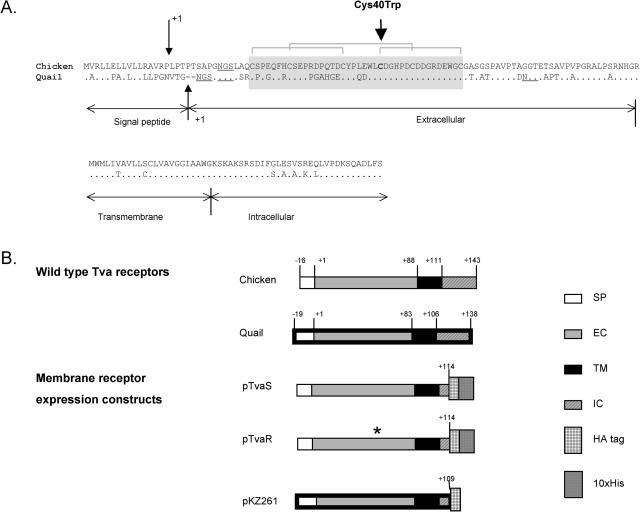
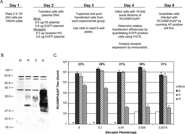
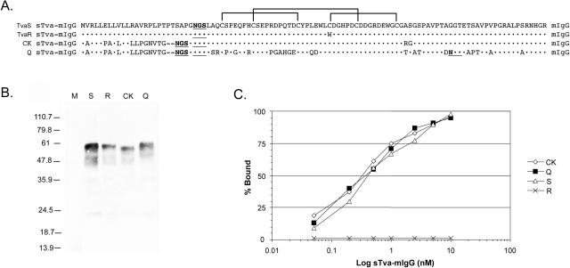
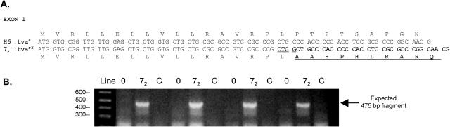
The subgroup A to E avian sarcoma and leukosis viruses (ASLVs) are highly related and are thought to have evolved from a common ancestor. These viruses use distinct cell surface proteins as receptors to gain entry into avian cells. Chickens have evolved resistance to infection by the ASLVs. We have identified the mutations responsible for the block to virus entry in chicken lines resistant to infection by subgroup A ASLVs [ASLV(A)]. The tva genetic locus determines the susceptibility of chicken cells to ASLV(A) viruses. In quail, the ASLV(A) susceptibility allele tva(s) encodes two forms of the Tva receptor; these proteins are translated from alternatively spliced mRNAs. The normal cellular function of the Tva receptor is unknown; however, the extracellular domain contains a 40-amino-acid, cysteine-rich region that is homologous to the ligand binding region of the low-density lipoprotein receptor (LDLR) proteins. The chicken tva(s) cDNAs had not yet been fully characterized; we cloned the chicken tva cDNAs from two lines of subgroup A-susceptible chickens, line H6 and line 0. Two types of chicken tva(s) cDNAs were obtained. These cDNAs encode a longer and shorter form of the Tva receptor homologous to the Tva forms in quail. Two different defects were identified in cDNAs cloned from two different ASLV(A)-resistant inbred chickens, line C and line 7(2). Line C tva(r) contains a single base pair substitution, resulting in a cysteine-to-tryptophan change in the LDLR-like region of Tva. This mutation drastically reduces the binding affinity of Tva(R) for the ASLV(A) envelope glycoproteins. Line 7(2) tva(r2) contains a 4-bp insertion in exon 1 that causes a change in the reading frame, which blocks expression of the Tva receptor.


Figures

FIG. 1.
FIG. 1.
Two alternatively spliced Tva transcripts are present in ASLV(A)-sensitive and -resistant chicken cells. RNA samples were prepared from CEFs of inbred lines H6 (sensitive) and C (resistant) and subjected to RT and PCR amplification. The primers used span the entire predicted Tva coding region and yield cDNA products from the longer and shorter alternatively spliced RNAs of 569 and 420 bp, respectively. The nucleotide sequences of the gel-purified cDNA products were determined directly. The nucleotide and deduced amino acid sequences of the coding regions of the long and short chicken Tva cDNAs are shown. Identical nucleotides are represented by dashes, and vertical lines mark the exon boundaries. The 40-amino-acid LDLR homologous cysteine-rich sequence (LDL-A) is highlighted in a shaded box. The putative transmembrane segments are highlighted in open boxes. We assume that the shorter form of Tva is attached to the cell surface via a glycosyl phosphatidylinositol linkage. The codon containing the single nucleotide difference between the line H6 and line C Tva cDNAs that results in the Cys40Trp mutation is labeled line C.
FIG. 2.
FIG. 2.
The nucleotide and putative amino acid sequences of the chicken tva locus of line H6. The genomic DNA region of tva was amplified by PCR, and the nucleotide sequence was determined. Each exon is shown in bold with the deduced amino acid sequence. The alternate exon of the long cDNA is highlighted with a shaded box. The nucleotide sequence of the tva introns and flanking regions are shown, and their boundaries are underlined and numbered. The codon containing the single nucleotide difference between the tva cDNAs from line H6 and line C that results in the Cys40Trp mutation is labeled line C.
FIG. 3.
FIG. 3.
The chicken and quail Tva receptors and the expression constructs. (A) Comparison of the deduced amino acid sequences of the chicken and quail Tva receptors. Identical amino acids are denoted by dots, and gaps are indicated by dashes. Only the longer forms of both receptors are shown. The quail receptor is predicted to have a signal peptide of 19 residues, followed by an extracellular domain of 83 residues which includes the LDL-A module, marked by a shaded box (9). The chicken Tva receptor is predicted to have a leader peptide of 16 amino acids (32) followed by an extracellular domain of 88 residues. The predicted cleavage sites of the leader peptidase are marked by small vertical arrows, and the amino acid numbering starts with the first residue (+1) of the mature chicken and quail proteins generated by the cleavage. There is considerable similarity in the sequences of the Tva proteins, and the extracellular, transmembrane, and intracellular regions of the chicken receptor are assumed to correspond to the regions in the quail Tva protein. The transmembrane and cytoplasmic tail regions of both proteins consist of 23 and 32 amino acids, respectively. The three disulfide bonds in the LDL-A module are indicated by brackets, and the potential N-linked glycosylation sites are underlined. The large vertical arrow marks the site of the mutation in line C, which involves Cys40 in chicken Tva (corresponding to Cys35 in quail Tva). (B) Schematic diagram of the Tva receptors and expression constructs used in these studies. Only the longer forms of the chicken and quail Tva receptors are shown; their extracellular, transmembrane, and cytoplasmic regions were included in the expression constructs. The construct pKZ261 was described previously (55). The constructs pTvaR and pTvaS were generated specifically for these experiments and are described in Materials and Methods. The horizontal bars with thin and thick black boundaries denote sequences of chicken and quail origin, respectively. The numbers indicate the position of amino acid residues in mature proteins; signal peptide residues have negative numbers. The asterisk indicates the presence of the Cys40Trp mutation in the pTvaR construct. SP, signal peptide; EC, extracellular domain; TM, transmembrane domain; IC, intracellular domain; HA, epitope tag from influenza virus HA protein; His, histidine residue tag.
FIG. 4.
FIG. 4.
The different Tva receptors confer different susceptibilities to ASLV(A) infection. (A) Schematic representation of the experimental approach. (B) Western immunoblot analysis of the levels of Tva receptor expressed by 293 cells transfected with 8 μg of receptor plasmid DNAs. Clarified cell lysates were made, the proteins were separated by SDS-12% PAGE and transferred to nitrocellulose, the filter was probed with anti-HA monoclonal antibody 12CA5, and the bound protein complexes were visualized by chemiluminescence. Molecular sizes (in kilodaltons) are given on the left. Lanes: M, mock; Q, quail Tva950; S, chicken TvaS; R, chicken TvaR. (C) 293 cells were transfected with 8.8 μg of plasmid DNA that contained different amounts of a plasmid encoding a Tva receptor. Transfected 293 cells expressing the different Tva receptors, quail Tva950 (Q), chicken TvaS (S), chicken TvaR (R), or mock (M), were challenged with 10-fold serial dilutions of RCASBP(A)AP and the titers were determined by AP assay. The asterisk indicates that the titer was below the limit of detection, 1 IFU/ml. The transfection efficiencies for each experimental group are shown above the titers as an average percentage. The results are averages of triplicate experiments. Error bars show standard deviations.
FIG. 5.
FIG. 5.
Binding affinity of the Tva receptors for ASLV(A) envelope glycoproteins. (A) Comparison of the signal peptides and extracellular domains of the quail Tva receptor (Q), the previously published quail/chicken Tva receptor (CK), the chicken TvaS receptor, and the chicken TvaR receptor used in the soluble Tva receptor constructs. Tva amino acids identical to the TvaS receptor are indicated (•). The three disulfide bonds are indicated by brackets. (B) Western immunoblot analysis of the sTva-mIgG proteins. The sTva-mIgG proteins were immunoprecipitated with goat anti-mouse IgG-agarose beads from clarified supernatants collected from the clonal DF-1 producer lines, denatured, separated by SDS-12% PAGE, and transferred to nitrocellulose. The filter was probed with peroxidase-conjugated goat anti-mouse IgG, and the bound protein-antibody complexes were visualized by chemiluminescence using Kodak X-Omat film. M, mock supernatant; S, chicken TvaS sTva-mIgG; R, chicken TvaR sTva-mIgG; CK, chicken sTva-mIgG; Q, quail sTva-mIgG. Molecular sizes (in kilodaltons) are given on the left. (C) Binding of the sTva-mIgG proteins to ASLV(A) glycoproteins. Uninfected DF-1 cells and DF-1 cells infected with wild-type RCASBP(A)AP virus were fixed with paraformaldehyde and incubated with different amounts of a sTva-mIgG protein and the envelope glycoprotein/sTva-mIgG complexes bound to goat anti-mouse Ig antibody linked to phycoerythrin. The amount of phycoerythrin bound to the cells was determined by FACS, and the maximum fluorescence was estimated (see Materials and Methods). The data were plotted as percent maximum fluorescence bound versus sTva-mIgG concentration. The values shown are averages of duplicate experiments.
FIG. 6.
FIG. 6.
The tvar2 gene of chicken Line 72 contains a 4-nucleotide insertion. (A) The nucleotide sequence and deduced amino acid sequence of exon 1 of the tvas gene of the ASLV(A)-susceptible line H6 (H6:tvas) and the tvar2 gene of ASLV(A)-resistant line 72 (72:tvar2). The 4-nucleotide insertion and altered amino acid sequence of line 72 are highlighted in bold. (B) A PCR assay was developed to specifically detect the 4-nucleotide insertion in the tvar2 gene of line 72 genomic DNA. The picture shows a gel electrophoresis of DNAs produced by PCR amplification of four samples each of line 0 (0), line 72 (72), and line Rh-C (C) genomic DNA isolated from CEFs separated on a 2% agarose gel and visualized with ethidium bromide. The sizes of the DNA markers are shown on the left in base pairs.
FIG. 7.
FIG. 7.
Maps of the regions encompassing syntenic genes in human, mouse, and chicken. Horizontal bars symbolize chromosomes; the organism name is given on the left, the chromosome number is on the right, and the gene symbols are on the top. The symbols of orthologous human genes are indicated in parentheses. The chicken genes represented by database entries ChEST112m16 and BX932614 showed the highest homology on a whole-genome level to human genes RPS28 and NDUFA7, respectively, as determined by the BLAST program (4). The directions of transcription of the genes are shown with arrows; the numbers between arrows indicate intergenic distances (in kilobases). The drawing is not to scale.

Similar articles

Cited by

References

    1. Adkins, H. B., S. C. Blacklow, and J. A. T. Young. 2001. Two functionally distinct forms of a retroviral receptor explain the nonreciprocal receptor interference among subgroups B, D, and E avian leukosis viruses. J. Virol. 75:3520-3526. - PMC - PubMed
    1. Adkins, H. B., J. Brojatsch, J. Naughton, M. M. Rolls, J. M. Pesola, and J. A. T. Young. 1997. Identification of a cellular receptor for subgroup E avian leukosis virus. Proc. Natl. Acad. Sci. USA 94:11617-11622. - PMC - PubMed
    1. Adkins, H. B., J. Brojatsch, and J. A. T. Young. 2000. Identification and characterization of a shared TNFR-related receptor for subgroup B, D and E avian leukosis viruses reveal cysteine residues required specifically for subgroup E viral entry. J. Virol. 74:3572-3578. - PMC - PubMed
    1. Altschul, S. F., T. L. Madden, A. A. Schaffer, J. Zhang, Z. Zhang, W. Miller, and D. J. Lipman. 1997. Gapped BLAST and PSI-BLAST: a new generation of protein database search programs. Nucleic Acid Res. 25:3389-3402. - PMC - PubMed
    1. Bacon, L. D., H. D. Hunt, and H. H. Cheng. 2000. A review of the development of chicken lines to resolve genes determining resistance to diseases. Poultry Sci. 79:1082-1093. - PubMed

Publication types

MeSH terms

LinkOut - more resources