. 2004 Dec;78(24):13501-11.
doi: 10.1128/JVI.78.24.13501-13511.2004.

Assembly protein precursor (pUL80.5 homolog) of simian cytomegalovirus is phosphorylated at a glycogen synthase kinase 3 site and its downstream "priming" site: phosphorylation affects interactions of protein with itself and with major capsid protein

Affiliations

Assembly protein precursor (pUL80.5 homolog) of simian cytomegalovirus is phosphorylated at a glycogen synthase kinase 3 site and its downstream "priming" site: phosphorylation affects interactions of protein with itself and with major capsid protein

Rebecca J Casaday et al. J Virol. 2004 Dec.

Abstract

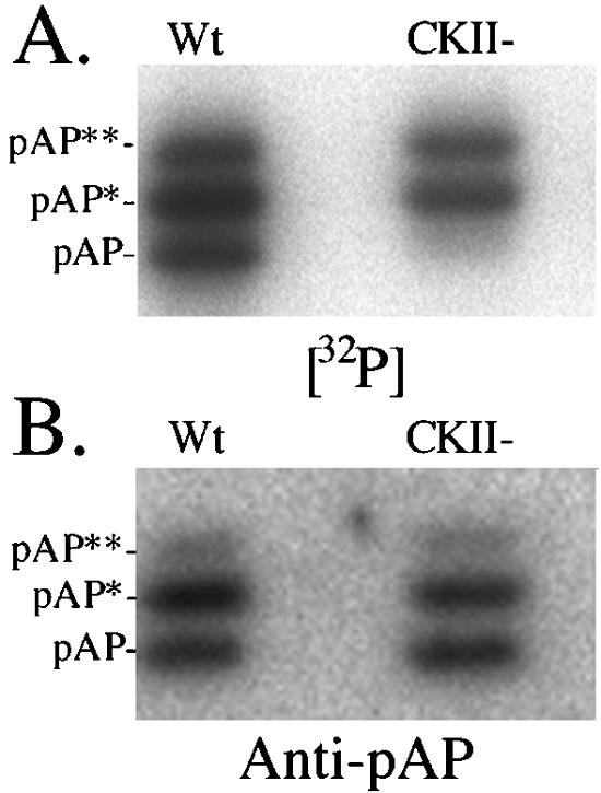
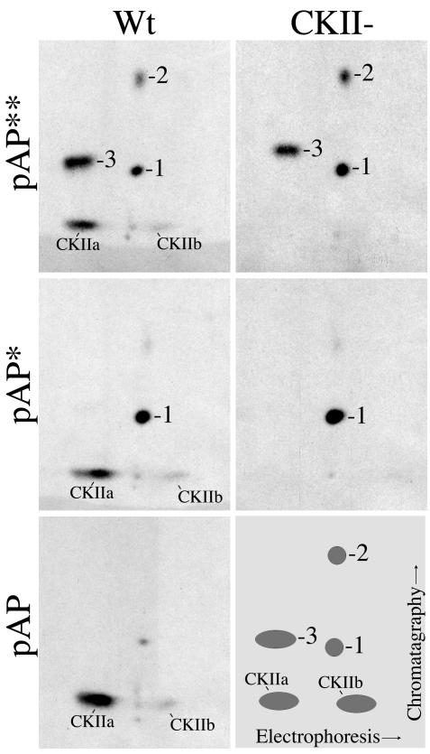
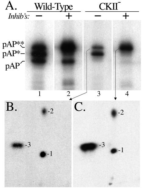
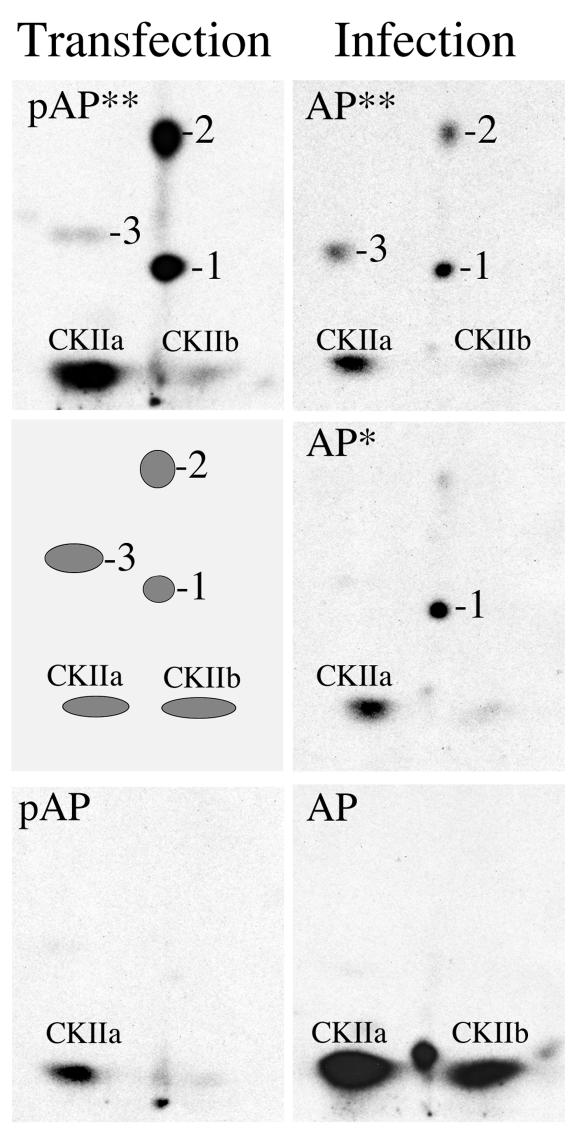
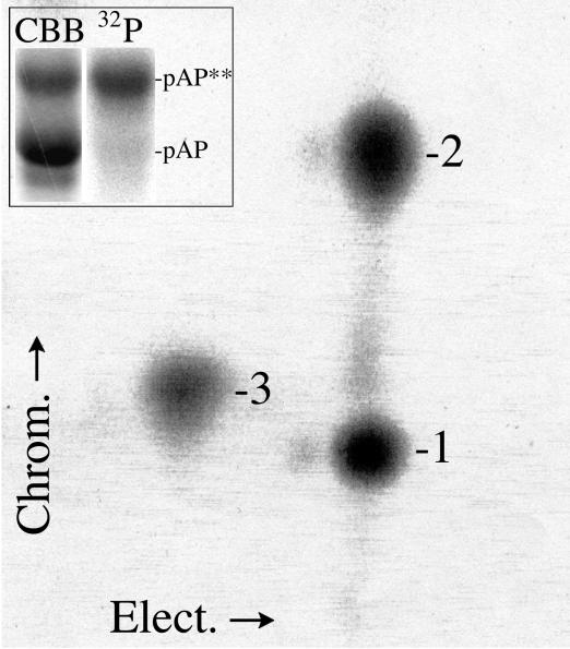
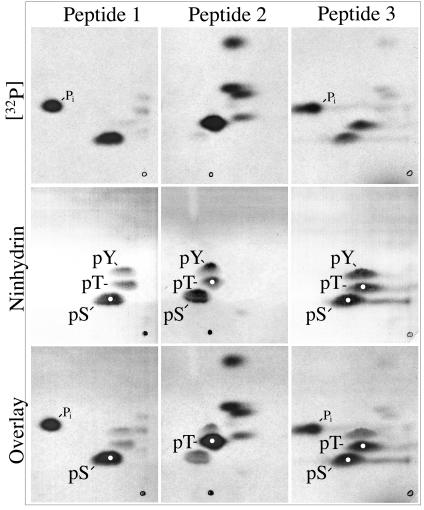
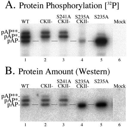
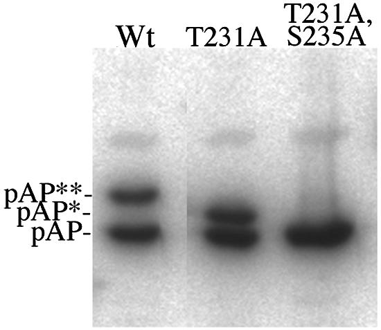
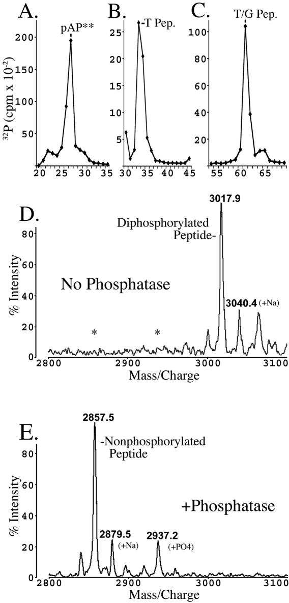
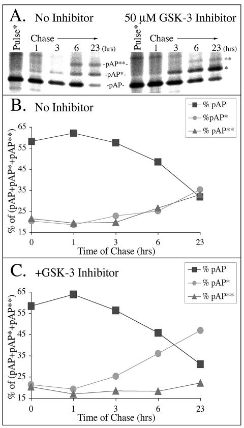
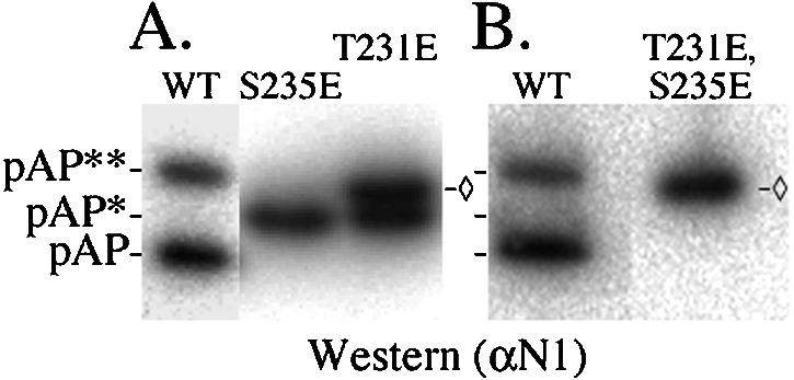
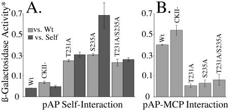
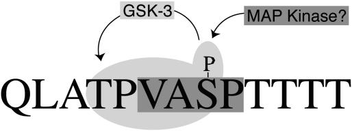
Capsid assembly among the herpes-group viruses is coordinated by two related scaffolding proteins. In cytomegalovirus (CMV), the main scaffolding constituent is called the assembly protein precursor (pAP). Like its homologs in other herpesviruses, pAP is modified by proteolytic cleavage and phosphorylation. Cleavage is essential for capsid maturation and production of infectious virus, but the role of phosphorylation is undetermined. As a first step in evaluating the significance of this modification, we have identified the specific sites of phosphorylation in the simian CMV pAP. Two were established previously to be adjacent serines (Ser156 and Ser157) in a casein kinase II consensus sequence. The remaining two, identified here as Thr231 and Ser235, are within consensus sequences for glycogen synthase kinase 3 (GSK-3) and mitogen-activated protein kinase, respectively. Consistent with Thr231 being a GSK-3 substrate, its phosphorylation required a downstream "priming" phosphate (i.e., Ser235) and was reduced by a GSK-3-specific inhibitor. Phosphorylation of Ser235 converts pAP to an electrophoretically slower-mobility isoform, pAP*; subsequent phosphorylation of pAP* at Thr231 converts pAP* to a still-slower isoform, pAP**. The mobility shift to pAP* was mimicked by substituting an acidic amino acid for either Thr231 or Ser235, but the shift to pAP** required that both positions be phosphorylated. Glu did not substitute for pSer235 in promoting phosphorylation of Thr231. We suggest that phosphorylation of Thr231 and Ser235 causes charge-driven conformational changes in pAP, and we demonstrate that preventing these modifications alters interactions of pAP with itself and with major capsid protein, suggesting a functional significance.


Figures

FIG. 1.
FIG. 1.
SCMV pAP resolves into three electrophoretic mobility isoforms during SDS-PAGE. Wild-type (Wt) and CKII precursor assembly proteins were expressed, 32P labeled in transfected cells, recovered by immunoprecipitation in the absence of phosphatase inhibitors, and subjected to SDS-PAGE and electrotransfer to PVDF Immobilon membranes (Millipore, Bedford, Mass.). (A) phosphorimage of the dried membrane showing the 32P-labeled radioactivity of each isoform. (B) Following radiodecay of the 32P (18 half-lives; undetected by phosphorimager), the membrane was probed with anti-N1 and then 125I-labeled protein A and phosphorimaged to determine the relative amount of each isoform.
FIG. 2.
FIG. 2.
pAP isoforms have different phosphopeptide patterns. Wild-type (Wt) and CKII precursor assembly proteins were expressed and 32P labeled in transfected cells, and the isoforms were immunoprecipitated in the absence of phosphatase inhibitors, resolved by SDS-PAGE, and subjected to pronase cleavage and peptide analysis as described in Materials and Methods. Shown here are fluorographic images of the resulting thin-layer plates. In place of an image for CKII pAP, which is not phosphorylated (e.g., Fig. 1A) (43), a schematic is presented showing the wild-type pAP** phosphopeptide designations (bottom right).
FIG. 3.
FIG. 3.
Phosphatase inhibitors increase the relative amount of pAP**. Wild-type and CKII precursor assembly proteins were expressed, 32P labeled in transfected cells, and recovered by immunoprecipitation without (−) or with (+) phosphatase inhibitors present as described in Materials and Methods and Results. Following SDS-PAGE and electrotransfer to an Immobilon membrane, an autoradiogram was made of the resulting membrane (A). The pAP** bands were cut from a second gel containing the same preparations, and their phosphopeptide patterns were compared as described in Materials and Methods. Fluorographic images prepared from the resulting thin-layer separations of phosphopeptides from pAP** recovered without (B) or with (C) phosphatase inhibitors are shown. Phosphopeptide numbering is the same as that in Fig. 2.
FIG. 4.
FIG. 4.
Phosphopeptide patterns of pAP isoforms from plasmid-transfected and AP isoforms from virus-infected cells are similar. Phosphopeptides were prepared from pAP isoforms immunoprecipitated from transfected cells (Transfection) and compared with those of AP isoforms recovered from infected cells (Infection), as described in Materials and Methods. Shown here are fluorographic images of the resulting thin-layer plates. With phosphatase inhibitors present, too little pAP* was recovered from transfected cells for peptide analysis; a schematic representation of the pAP** phosphopeptides is presented in its place. Phosphopeptide numbering is the same as that in Fig. 2.
FIG. 5.
FIG. 5.
pAP** expressed from rBV KC34 has a charasteristic pronase phosphopeptide pattern. 32P-labeled phosphopeptides were prepared from CKII pAP** expressed in insect cells and analyzed by 2-D separation, as described in Materials and Methods. The inset shows the starting protein preparation recovered by Ni-immobilized metal affinity chromatography after SDS-PAGE, followed by staining with Coomassie brilliant blue (CBB) or autoradiography (32P) of the same gel lane. Shown here is a phosphorimage of the resulting phosphopeptide distribution. Phosphopeptides numbering is the same as that in Fig. 2.
FIG. 6.
FIG. 6.
Identification of phosphorylated amino acids in pronase-derived phosphopeptides 1, 2, and 3 from pAP isoforms. 32P-labeled phosphopeptides from rBV KC34-expressed pAP** were prepared, hydrolyzed, and subjected to amino acid analyses as described in Materials and Methods. Shown here are fluorographic images prepared from the resulting thin-layer plates (top); direct images of the same plates sprayed with ninhydrin to detect the pThr (pT), pSer (pS), and pTyr (pY) standards added to each sample (middle); and composite images of the superimposed fluorographic and ninhydrin images (bottom). White dots indicate phosphoamino acid coincident with radioactivity in that sample. Two hydrolysates were analyzed per plate. The sample origin is seen as a spot or small circle at the bottom. Electrophoresis was left (+) to right (−), followed by ascending chromatography.
FIG. 7.
FIG. 7.
Patterns of pAP mobility isoforms for wild-type and mutant forms of the proteins. (A) 32P-labeled proteins were immunoprecipitated in the absence of phosphatase inhibitors from cells expressing the wild type (WT) or the indicated mutations and subjected to SDS-PAGE followed by electrotransfer to Immobilon membranes and phosphorimaging. (B) Once 32P radioactivity decayed below detectability by phosphorimaging, the membrane was subjected to Western immunoassay with anti-N1 and 125I-labeled protein A and phosphorimaged. Preparation of mock-transfected cells (Mock) was carried out in a noncontiguous lane and has been transposed in this collage. Lines between the lanes indicate the position of pAP.
FIG. 8.
FIG. 8.
Mutation of Thr231 eliminates only pAP**. Wild-type pAP (Wt) and the T231A and T231A/S235A mutations were expressed in transfected cells, harvested into 4× protein sample buffer at 70°C, and analyzed by Western immunoassay with anti-N1 and 125I-labeled protein A, following SDS-PAGE. Shown here is a phosphorimage of the resulting membrane. Preparation of the wild type took place in a noncontiguous lane and has been transposed in this collage.
FIG. 9.
FIG. 9.
Mass spectrometry confirms presence of two phosphates on peptide containing Thr231 and Ser235. The Gln217-Glu245 trypsin/GluC peptide was prepared from 32P-labeled pAP** expressed from rBV KC34, as described in Materials and Methods and Results. The top panels show the radioactivity patterns resulting from RP-HPLC separation of the protein, pAP** (A), the tryptic peptide (T Pep.) (B), and the double-cut trypsin/GluC peptide (T/G Pep.) (C). The resulting peptide was subjected to MALDI-TOF without further treatment (D) or following treatment with calf alkaline phosphatase (E). Asterisks in panel D indicate predicted positions of nonphosphorylated and monophosphorylated peptide (E). Ions at 3040.4 (D) and 2879.5 (E) correspond to sodiated forms of the diphosphorylated and nonphosphorylated peptide, respectively.
FIG. 10.
FIG. 10.
GSK-3 inhibitor selectively reduces relative amount of pAP**. [35S]methionine-labeled transfected-cell proteins were immunoprecipitated with anti-C1 and subjected to SDS-PAGE and phosphorimaging (A). Phosphatase inhibitors (catalogue no. 524625, Calbiochem) but no protease inhibitors were added to the lysis buffer, antiserum, and all immunoprecipitation solutions. In preparation for SDS-PAGE, proteins were solubilized in a solution containing six parts NuPAGE lithium dodecyl sulfate sample buffer (catalogue no. NP0007; Invitrogen) and four parts 1 M dithiothreitol. Total radioactivity in the three pAP isoforms was measured for each time point, and the relative percentage of the total was calculated for each isoform. Shown here are the changes in the percentage of the total for each isoform from nontreated cells (B) and cells treated with GSK-3 inhibitor (C).
FIG. 11.
FIG. 11.
Substituting glutamic acid for Thr231 or Ser235 produces electrophoretically slower isomers of pAP. Cells were transfected to express pAP mutation T231E, S235E, or T231E/S235E. Four days later, the cells were harvested into 4× protein sample buffer at 70°C and subjected to SDS-PAGE, followed by Western immunoassay with anti-N1 and phosphorimaging, as described in Materials and Methods. Shown here is a phosphorimage of the resulting Immobilon membrane. ◊, position of T231E/S235E mutant isoform, as distinct from pAP**.
FIG. 12.
FIG. 12.
Phosphorylation mutants show stronger interactions with self, but weaker interactions with MCP. Yeast GAL4 two-hybrid assays were done as described in Materials and Methods and Results to test the interaction of pAP phosphorylation mutants with themselves, wild-type pAP, and MCP. Data shown are averages ± standard errors (as described in Materials and Methods) from three separate experiments testing self-interaction (A) and three experiments testing interaction with MCP (B). Self interactions were between heterologous pairs, i.e., wild-type (Wt) pAP with mutant pAP, where lighter results indicate the left-hand bar for each mutant, or between homologous pairs, where darker areas indicate the right-hand bar for each protein. Wild-type pAP had no heterologous pair (A); the values for T231A/S235A versus MCP (B) were derived from two, rather than three, separate experiments. *, β-Galactosidase acitivity was calculated in Miller units (A420 × 1,000)/(min × 0.1 × concentration factor × optical density at 600 nm) (35).
FIG. 13.
FIG. 13.
Model showing the location of Thr231 and Ser235 within the pAP sequence containing consensus GSK-3 (light gray; includes GSK-3 priming phosphate) and MAP kinase (darker gray) phosphorylation sites.

Similar articles

Cited by

References

    1. Ausubel, F. M., R. Brent, R. E. Kingston, D. D. Moore, J. G. Seidman, J. A. Smith, and K. Struhl. 1990. Current protocols in molecular biology. John Wiley and Sons, New York, N.Y.
    1. Beaudet-Miller, M., R. Zhang, J. Durkin, W. Gibson, A. D. Kwong, and Z. Hong. 1996. Viral specific interaction between the human cytomegalovirus major capsid protein and the C-terminus of the precursor assembly protein. J. Virol. 70:8081-8088. - PMC - PubMed
    1. Chevray, P. M., and D. Nathans. 1992. Protein interaction cloning in yeast: identification of mammalian proteins that react with the leucine zipper of Jun. Proc. Natl. Acad. Sci. USA 89:5789-5793. - PMC - PubMed
    1. Coghlan, M. P., A. A. Culbert, D. A. Cross, S. L. Corcoran, J. W. Yates, N. J. Pearce, O. L. Rausch, G. J. Murphy, P. S. Carter, L. R. Cox, D. Mills, M. J. Brown, D. Haigh, R. W. Ward, D. G. Smith, K. J. Murray, A. D. Reith, and J. C. Holder. 2000. Selective small molecule inhibitors of glycogen synthase kinase-3 modulate glycogen metabolism and gene transcription. Chem. Biol. 7:793-803. - PubMed
    1. Desai, P., and S. Person. 1996. Molecular interactions between the HSV-1 capsid proteins as measured by the yeast two-hybrid system. Virology 220:516-521. - PubMed

Publication types

LinkOut - more resources