. 2004 Dec;78(24):13573-81.
doi: 10.1128/JVI.78.24.13573-13581.2004.

Furin-mediated cleavage of the feline foamy virus Env leader protein

Affiliations

Furin-mediated cleavage of the feline foamy virus Env leader protein

Verena Geiselhart et al. J Virol. 2004 Dec.

Abstract

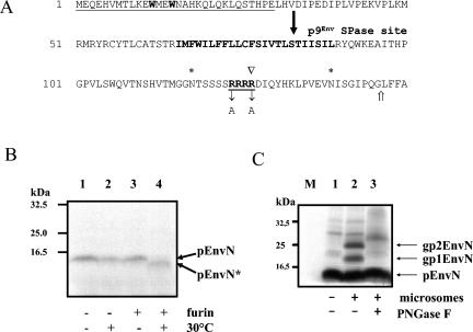
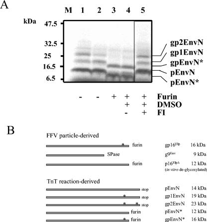
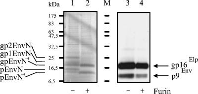
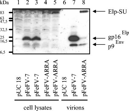
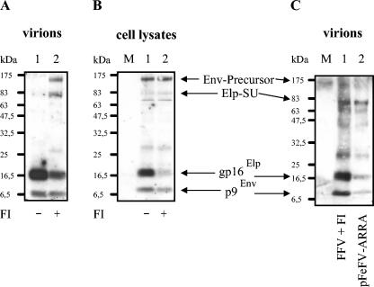
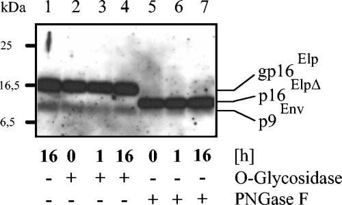
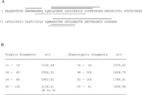
The molecular biology of spuma or foamy retroviruses is different from that of the other members of the Retroviridae. Among the distinguishing features, the N-terminal domain of the foamy virus Env glycoprotein, the 16-kDa Env leader protein Elp, is a component of released, infectious virions and is required for particle budding. The transmembrane protein Elp specifically interacts with N-terminal Gag sequences during morphogenesis. In this study, we investigate the mechanism of Elp release from the Env precursor protein. By a combination of genetic, biochemical, and biophysical methods, we show that the feline foamy virus (FFV) Elp is released by a cellular furin-like protease, most likely furin itself, generating an Elp protein consisting of 127 amino acid residues. The cleavage site fully conforms to the rules for an optimal furin site. Proteolytic processing at the furin cleavage site is required for full infectivity of FFV. However, utilization of other furin proteases and/or cleavage at a suboptimal signal peptidase cleavage site can partially rescue virus viability. In addition, we show that FFV Elp carries an N-linked oligosaccharide that is not conserved among the known foamy viruses.


Figures

FIG. 1.
FIG. 1.
Identification of a furin cleavage site between FFV Env residues 127 and 128 by using in vitro-synthesized pEnvN protein. (A) Sequence of the N-terminal 150 amino acid residues (in single-letter code) of FFV Env with the following features marked: residues W12 and W15 required for Gag-Env interaction in boldface type; the region against which the Elp serum is directed is underlined; the hydrophobic TM anchor is marked in boldface type; potential N-glycosylation sites are marked by asterisks above the sequence; the RRRR motif specifying a furin cleavage site (open arrowhead) is underlined and in boldface type, with the Arg-Ala exchanges of the mutant pFeFV-ARRA indicated below the sequence; the end of the in vitro-synthesized N-terminal Env protein pEnvN is marked by an open arrow. The solid arrow marks a potential SPase cleavage site (11, 18, 33). (B) To characterize in vitro-synthesized pEnvN, TNT reactions were run with pBl-FFV-EnvN template DNA, supplemented with furin buffer, and incubated at 30°C for 120 min in the absence (−) and presence (+) of furin, as indicated below the autoradiogram. Samples in lanes 1 and 3 were taken before the reactions were started. The reaction products were analyzed by gel electrophoresis and autoradiography. The in vitro-synthesized pEnvN protein with an apparent molecular mass of 14 kDa is marked by an arrow. Incubation with furin (lane 4) resulted in the disappearance of the 14-kDa band and the appearance of the 12-kDa pEnvN* furin cleavage product (arrow). (C) In vitro glycosylation of pEnvN. As above, pEnvN was in vitro synthesized in the absence (lane 1) or presence of microsomes, as indicated below the lanes. The reaction products were separated by PAGE and visualized by autoradiography. In all reactions, the primary translation product pEnvN is clearly present. The addition of microsomes induced two specific bands of about 19 (gp1EnvN) and 23 (gp2EnvN) kDa (lane 2) that were both susceptible to digestion with the N-glycosidase PNGaseF (lanes 3). The positions of marker proteins separated in parallel are shown to the left.
FIG. 2.
FIG. 2.
In vitro glycosylation and furin processing of pEnvN and schematic presentation of reaction products. (A) pEnvN was in vitro synthesized in the presence of microsomes. After completion of the TNT reaction, an aliquot was directly mixed with gel loading buffer (lane 1), the rest was supplemented with furin reaction buffer, and aliquots were incubated for 4 h at 30°C with (+) 0.1 U of furin (lanes 3 to 5) or without (−) furin (lane 2). As a control for solvent effects, the sample in lane 4 was supplemented with DMSO, the sample in lane 5 contained 620-μg/ml FI in DMSO, and lane 3 did not contain DMSO or FI. Reaction products were separated by PAGE and visualized by autoradiography. In the reaction mixtures in lanes 1 and 2, the primary translation product pEnvN and the glycosylated forms gp1EnvN and gp2EnvN were clearly present. The addition of furin (lanes 3 and 4) resulted in the disappearance of these bands and the appearance of two new bands of 16 (gpEnvN*) and 12 (pEnvN*) kDa that were both repressed by FI (lane 5). The positions of marker proteins separated in parallel are shown on the left. (B) Comparative schematic presentation of FFV virion-derived Elp and p9Env proteins (top) and the pEnvN-derived proteins generated in TNT reactions (bottom). The sizes of the different proteins starting at Env residue 1 are schematically given by the size of the bars. The C termini were generated either by furin, SPase, or a stop codon in the TNT template DNA, as indicated. Protein modifications by glycosylation are marked by asterisks. For each protein, the name and its apparent molecular mass are given. Note that gp16Elp and p16ElpΔ are identical to gpEnvN* and pEnvN*, respectively.
FIG. 3.
FIG. 3.
Comigration of virion-derived and in vitro-synthesized Elp. pEnvN was in vitro synthesized in the TNT system in the presence of microsomes, and an aliquot of the reaction product was subsequently digested with furin (lanes 1 and 2, indicated below the lanes). In parallel, released FFV particles were enriched from supernatants of FFV-infected cells by sedimentation through a sucrose cushion. An aliquot of the FFV particles was digested with furin in parallel with the in vitro-synthesized pEnvN proteins (lanes 3 and 4, indicated below the lanes). The reaction products were analyzed on a single gel separated by a lane loaded with prestained molecular mass markers (lane M). Lanes 1 and 2 together with a part of the marker lane were dried, and the labeled reaction products were visualized by autoradiography (marked on the left). The remainder of the gel (part of lane M and lanes 3 and 4) was subjected to immunoblotting with the Elp serum and detected by ECL (marked on the right). The alignment of the images and comparisons with the marker proteins show that the in vitro-generated gpEnvN* perfectly comigrates with the mature virion-derived gp16Elp. The positions of marker proteins are given on the left. +, present; −, absent.
FIG. 4.
FIG. 4.
Analysis of Env proteins expressed from mutant pFeFV-ARRA. 293T cells were transfected in duplicate with plasmids pFeFV-7 (lanes 2 and 3) and pFeFV-ARRA (lanes 4 and 5). Cellular extracts were harvested 3 days posttransfection, and equal amounts of proteins were subjected to immunoblotting with the Elp-specific antiserum. In parallel, particles were concentrated from the pooled supernatants of the cells and analyzed on the same gel (lanes 7 and 8). pUC18-transfected cells served as controls (lanes 1 and 6). The Elp-related proteins p9Env and gp16Elp and the Elp-SU fusion protein were detected by ECL. Lanes 1 to 5 were intentionally overexposed to detect FFV Elp, p9Env, and the Elp-SU fusion protein.
FIG. 5.
FIG. 5.
Inhibition of Elp processing by the FI dec-RVKR-cmk. FFV-infected CRFK cells were incubated in the presence (+) or absence (−) of 7.4 μg of the FI dec-RVKR-cmk/ml as described in Materials and Methods between 4 and 72 h postinfection. Three days postinfection, the cells and supernatants were harvested. (A) Released particles were enriched from the cell culture supernatants and analyzed by immunoblotting with the Elp antiserum. Without FI, the mature Elp and the 9-kDa SP were detectable. With FI, the unprocessed Env precursor and the 83-kDa Elp-SU protein became visible, whereas only small amounts of the mature Elp were detectable. (B) In the cell-associated antigen, the amount of Elp was significantly reduced by FI. (C) The FFV virion-specific 83-kDa Elp-specific protein generated in the presence of FI (lane 1) and the 83-kDa Elp-specific protein of pFeFV-ARRA-derived particles (lane 2, compare to Fig. 4) comigrate when analyzed by immunoblotting on adjacent gel lanes.
FIG. 6.
FIG. 6.
Deglycosylation of virion-derived Elp. FFV virions were enriched by sedimentation through sucrose, and the viral proteins were denatured for glycosidase digestion as described in Materials and Methods. Aliquots were incubated for 16 h without (−) enzyme (lane 1) and for 0, 1, and 16 h with (+) O-glycosidase (lanes 2 to 4) or PNGaseF (lanes 5 to 7). The reaction products were detected by immunoblotting with the FFV Elp antiserum. The positions of molecular mass markers are marked on the left; the names of the detected FFV proteins are given on the right. Due to the high enzyme concentration used, PNGaseF cleavage had already occurred in the 0-h samples.
FIG. 7.
FIG. 7.
MALDI-TOF (MS) analysis of tryptic and chymotryptic fragments of FFV virion-derived Elp. FFV Elp was purified from FFV particles as described and used for peptide mass fingerprinting to define the C terminus of Elp. (A) The complete sequence of FFV Elp is given with a schematic presentation of the identified tryptic (solid lines above the sequence) and chymotryptic (dashed lines below the sequence) peptide fragments. (B) Presentation of the biophysical features (m/z values) of the Elp-derived tryptic (left column) and chymotryptic (right column) fragments identified by MALDI-TOF (MS) referring to the amino acid positions within Env/Elp. The deglycosylation-related N-to-D modification in the tryptic fragment 95 to 124 is indicated.

Similar articles

Cited by

References

    1. Alke, A., A. Schwantes, K. Kido, M. Flötenmeyer, R. M. Flügel, and M. Löchelt. 2001. The bet gene of feline foamy virus is required for virus replication. Virology 287:310-320. - PubMed
    1. Alke, A., A. Schwantes, M. Zemba, R. M. Flügel, and M. Löchelt. 2000. Characterization of the humoral immune response and virus replication in cats experimentally infected with feline foamy virus. Virology 275:170-176. - PubMed
    1. Bansal, A., K. L. Shaw, B. H. Edwards, P. A. Goepfert, and M. J. Mulligan. 2000. Characterization of the R572T point mutant of a putative cleavage site in human foamy virus Env. J. Virol. 74:2949-2954. - PMC - PubMed
    1. Basak, A., M. Zhong, J. Munzer, M. Chretien, and N. Seidah. 2001. Implication of the proprotein convertases furin, PC5 and PC7 in the cleavage of surface glycoproteins of Hong Kong, Ebola and respiratory syncytial viruses: a comparative analysis with fluorogenic peptides. Biochem. J. 353:537-545. - PMC - PubMed
    1. Bornsen, K. 2000. Influence of salts, buffers, detergents, solvents, and matrices on MALDI-MS protein analysis in complex mixtures. Methods Mol. Biol. 146:387-404. - PubMed

Publication types

LinkOut - more resources