. 2004 Dec;78(24):13717-26.
doi: 10.1128/JVI.78.24.13717-13726.2004.

Protein kinase C-alpha activity is required for respiratory syncytial virus fusion to human bronchial epithelial cells

Affiliations

Protein kinase C-alpha activity is required for respiratory syncytial virus fusion to human bronchial epithelial cells

Homero San-Juan-Vergara et al. J Virol. 2004 Dec.

Abstract

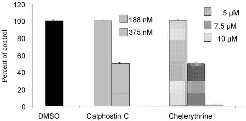
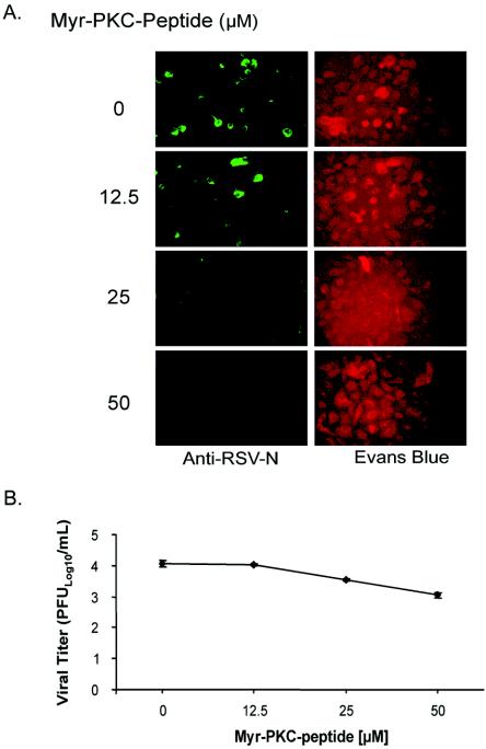
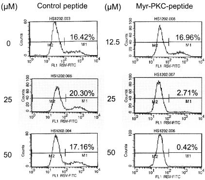
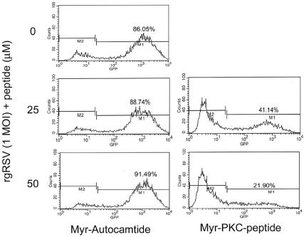
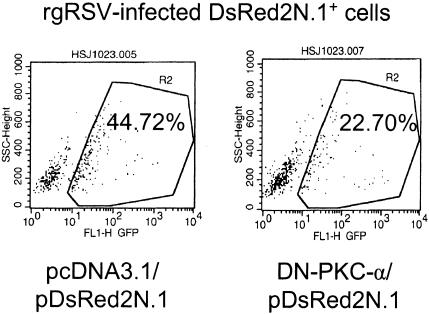
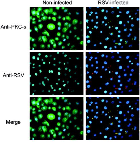
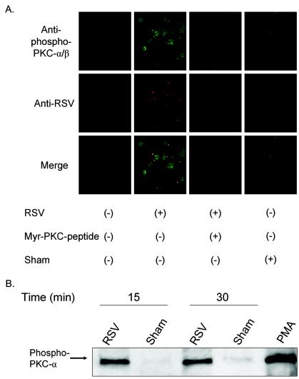
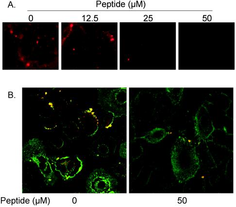
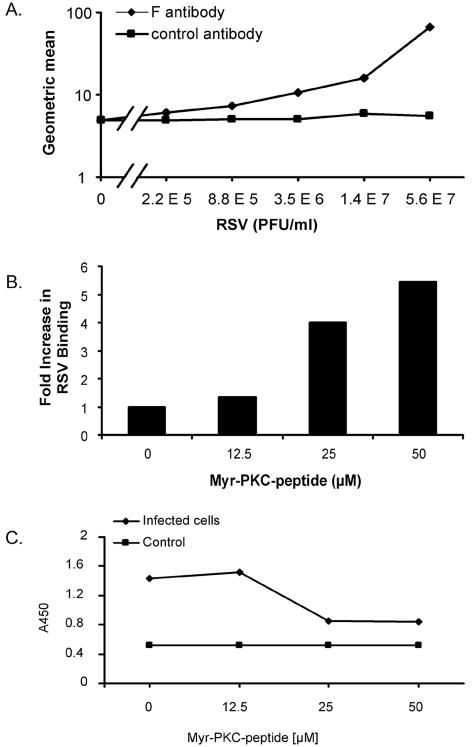
Respiratory syncytial virus (RSV) infection activates protein kinase C (PKC), but the precise PKC isoform(s) involved and its role(s) remain to be elucidated. On the basis of the activation kinetics of different signaling pathways and the effect of various PKC inhibitors, it was reasoned that PKC activation is important in the early stages of RSV infection, especially RSV fusion and/or replication. Herein, the role of PKC-alpha during the early stages of RSV infection in normal human bronchial epithelial cells is determined. The results show that the blocking of PKC-alpha activation by classical inhibitors, pseudosubstrate peptides, or the overexpression of dominant-negative mutants of PKC-alpha in these cells leads to significantly decreased RSV infection. RSV induces phosphorylation, activation, and cytoplasm-to-membrane translocation of PKC-alpha. Also, PKC-alpha colocalizes with virus particles and is required for RSV fusion to the cell membrane. Thus, PKC-alpha could provide a new pharmacological target for controlling RSV infection.


Figures

FIG. 1.
FIG. 1.
Inhibition of RSV infection of NHBE cells by PKC inhibitors. Confluent NHBE cells were treated with PKC inhibitors at different doses for 30 min before they were infected with RSV at an infectious dose of 1 MOI. The infection was allowed to proceed for 16 h, and infected cells were detected by single-cell immunofluorescence assays. The percentage of inhibition was calculated with respect to the control (dimethyl sulfoxide [DMSO]). The values are means ± standard deviations of three different experiments.
FIG. 2.
FIG. 2.
Inhibition of classical PKC isozymes impairs RSV infection and reduces virus production. (A) Confluent NHBE cells were treated with the myristoylated PKC-α/β pseudosubstrate peptide at the indicated concentrations for 30 min before being infected with RSV at an infectious dose of 1 MOI. The infection was allowed to proceed for 16 h. Infected cells were detected by single-cell immunofluorescence assays (magnification, ×200). (B) NHBE cells grown in a 12-well plate configuration were incubated with different concentrations of the myr-PKC peptide 30 min prior to being infected with 0.1 MOI of rgRSV. Infection was allowed to proceed for 60 h. Culture medium from each treatment condition was collected, and the viral titer was measured. The results are the averages ± standard deviations of three different experiments per experimental condition.
FIG. 3.
FIG. 3.
PKC-α/β pseudosubstrate peptide does not interfere with the RSV binding to NHBE cells. Confluent NHBE cells were treated with either the PKC pseudosubstrate peptide (control, nonmyristoylated) or the myristoylated PKC pseudosubstrate peptide (myr-PKC peptide) at the indicated concentrations for 30 min before being infected with RSV at an infectious dose of 1 MOI. The infection was allowed to proceed for 16 h. Cell culture monolayers were then detached by trypsin treatment, and single-cell suspensions were processed for analysis by fluorescence-activated cell sorter. The infected cells were detected by an FITC-labeled mouse monoclonal anti-RSV N protein antibody.
FIG. 4.
FIG. 4.
Myristoylated moiety did not block RSV binding to cells. Confluent NHBE cells were treated with either the myr-autocamtide or the myr-PKC peptide at the indicated concentrations for 30 min before being infected with rgRSV at an infectious dose of 1 MOI. The infection was allowed to proceed for 16 h. Cell culture monolayers were then detached by trypsin treatment, and single-cell suspensions were processed for analysis by fluorescence-activated cell sorter. Infected cells were detected by the fluorescence emitted by enhanced green fluorescent protein produced by rgRSV.
FIG. 5.
FIG. 5.
Expression of DN-PKC-α reduced the number of RSV-infected cells. Approximately 8 × 10⁵ NHBE cells grown in 6-well plates were transfected with 1 μg of either pDN-PKC-α or pcDNA3.1. Along with these plasmids, 100 ng of pDsRed2N1 was also cotransfected to visualize transfected cells by flow cytometry. Twenty-four hours later, cells were infected with rgRSV, and infection was indicated by the expression of enhanced green fluorescence protein. The percentage of infection in red fluorescent cells (infection in transfected cells) was then determined by flow cytometry.
FIG. 6.
FIG. 6.
PKC-α colocalizes with RSV at early stages of infection. Confluent NHBE cells grown on 8-well chamber slides were incubated with 20 MOI of RSV for 1 h at 4°C to allow viral binding synchronization. Slides were then exposed at 37°C for 10 min to allow virus penetration. Later, cells were processed for immunocytofluorescence. Cells were fixed with 4% paraformaldehyde, permeabilized with 0.1% saponin, and stained with mouse monoclonal anti-PKC-α antibody (green), goat polyclonal anti-RSV antibody (red), and DAPI (blue, nucleus staining). Fluorescence images (magnification, ×400) were taken by cooled camera device under respective dual-filter mode (either green-blue or red-blue) and triple-filter mode (merge).
FIG. 7.
FIG. 7.
Activated PKC-α colocalizes with RSV particles on the cell membrane. (A) Confluent NHBE cells grown on 8-well chamber slides were exposed to RSV at an infectious dose of 20 MOI for 10 min at 37°C following viral binding synchronization for 1 h at 4°C. As negative controls, cells were either pretreated with a PKC-α/β pseudosubstrate inhibitor peptide at 50 μM for 30 min before infection or exposed to a sham treatment (Centricon filtrate obtained from purified RSV). NHBE cells were fixed with 4% paraformaldehyde, permeabilized with 0.1% saponin, and stained with rabbit polyclonal anti-phospho-Thr-638 PKC-α antibody (green), goat polyclonal anti-RSV antibody (red), and DAPI (blue, nucleus staining). Confocal images (magnification, ×630) were taken by using laser excitation sources for Alexa-488 (green) or Alexa-555 (red) and assembled with Adobe Photoshop software version 7.01. (B) NHBE cells grown in T-25 flasks were infected with purified RSV at an infectious dose of 3 MOI for 15 or 30 min at 37°C following a viral binding synchronization step with incubation at 4°C for 1 h. Membrane fractions of each experimental condition were obtained, equal amounts of protein (8 μg) of the membrane fractions were analyzed by western blot, and the phospho-Thr-638 PKC-α was probed by the specific antibody.
FIG. 8.
FIG. 8.
(A) Inhibition of PKC-α activity blocks viral fusion. Confluent NHBE cells seeded onto 8-well chamber slides were preincubated with a PKC-α/β pseudosubstrate peptide for 30 min at the indicated concentrations before cells were exposed to octadecyl rhodamine B (R18)-labeled RSV (5,000 RSV particles/cell). The infection was allowed to proceed for 30 min at 37°C. After removal of the unattached virus, cells were imaged (magnification, ×200) by using a fluorescence microscope. (B) Myr-PKC-α/β pseudosubstrate peptide significantly reduces the number of viral cores inside NHBE cells. NHBE cells were infected on ice with purified RSV (20 MOI) and then incubated at 37°C for 1 h. Cells were then washed with ice-cold PBS containing FITC-WGA for the staining of the plasma membrane, as described above. Cells were again washed with ice-cold PBS, fixed with 3.7% paraformaldehyde, and permeabilized with 0.1% saponin. After washing with PBS, samples were treated for indirect immunofluorescence. Viral cores were probed by mouse monoclonal anti-RSV N protein antibodies and were revealed by rhodamine-labeled goat anti-mouse antibody. The pictures (magnification, ×630) represent single optical sections perpendicular to the z axis of a confocal microscope.
FIG. 9.
FIG. 9.
Quantitation of viral attachment and penetration. (A) NHBE cells in suspension were incubated on ice with serial fourfold dilutions of RSV to allow viral attachment. After 30 min, cells were washed three times with ice-cold staining buffer, and bound virus was detected with Zenon Alexa-488 anti-RSV F monoclonal antibody. Geometric mean fluorescence values were obtained and graphed. The curve for the isotype control antibody is also shown. (B) A total of 106 NHBE cells per ml in suspension were placed in the absence or presence of different concentrations of the myr-PKC peptide for 30 min at 37°C. Cells were washed with ice-cold basal medium, and 106 cells/ml were exposed to 1.4 × 10⁷ PFU of RSV/ml for 30 min on ice. Unbound virus was removed by washing in ice-cold staining buffer, and bound virus was detected by flow cytometry with Zenon Alexa-488 anti-RSV F monoclonal antibody. (C) NHBE cells grown on 6-well plates were incubated in the absence or presence of different concentrations of the myr-PKC peptide for 30 min before the cells were infected with 1.7 × 10⁶ PFU/well (approximately 5 MOI) for 2 h at 37°C. Cytoplasmic fractions were then isolated from each experimental condition, and a sandwich ELISA was conducted to probe the presence of RSV N protein in these fractions. TMB substrate was then added, and the plate was read at 450 nm (A450). The cytoplasmic fractions of noninfected cells were used as a control. These assays were duplicated with similar results.

References

    1. Bitko, V., and S. Barik. 1998. Persistent activation of RelA by respiratory syncytial virus involves protein kinase C, underphosphorylated IκBβ, and sequestration of protein phosphatase 2A by the viral phosphoprotein. J. Virol. 72:5610-5618. - PMC - PubMed
    1. Bitko, V., A. Velazquez, L. Yang, Y.-C. Yang, and S. Barik. 1997. Transcriptional induction of multiple cytokines by human respiratory syncytial virus requires activation of NF-κB and is inhibited by sodium salicylate and aspirin. Virology 232:369-378. - PubMed
    1. Brown, G., J. Aitken, H. W. Rixon, and R. J. Sugrue. 2002. Caveolin-1 is incorporated into mature respiratory syncytial virus particles during virus assembly on the surface of virus-infected cells. J. Gen. Virol. 83:611-621. - PubMed
    1. Brown, G., H. W. Rixon, and R. J. Sugrue. 2002. Respiratory syncytial virus assembly occurs in GM1-rich regions of the host-cell membrane and alters the cellular distribution of tyrosine phosphorylated caveolin-1. J. Gen. Virol. 83:1841-1850. - PubMed
    1. Budge, P. J., Y. Li, J. A. Beeler, and B. S. Graham. 2004. RhoA-derived peptide dimers share mechanistic properties with other polyanionic inhibitors of respiratory syncytial virus (RSV), including disruption of viral attachment and dependence on RSV G. J. Virol. 78:5015-5022. - PMC - PubMed

Publication types

MeSH terms

LinkOut - more resources