. 2004 Dec;78(24):13743-54.
doi: 10.1128/JVI.78.24.13743-13754.2004.

Protein transduction domains fused to virus receptors improve cellular virus uptake and enhance oncolysis by tumor-specific replicating vectors

Affiliations

Protein transduction domains fused to virus receptors improve cellular virus uptake and enhance oncolysis by tumor-specific replicating vectors

Florian Kühnel et al. J Virol. 2004 Dec.

Abstract

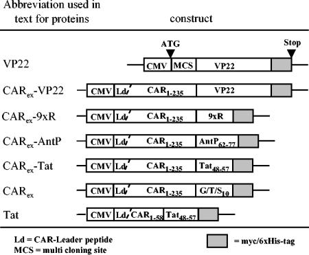
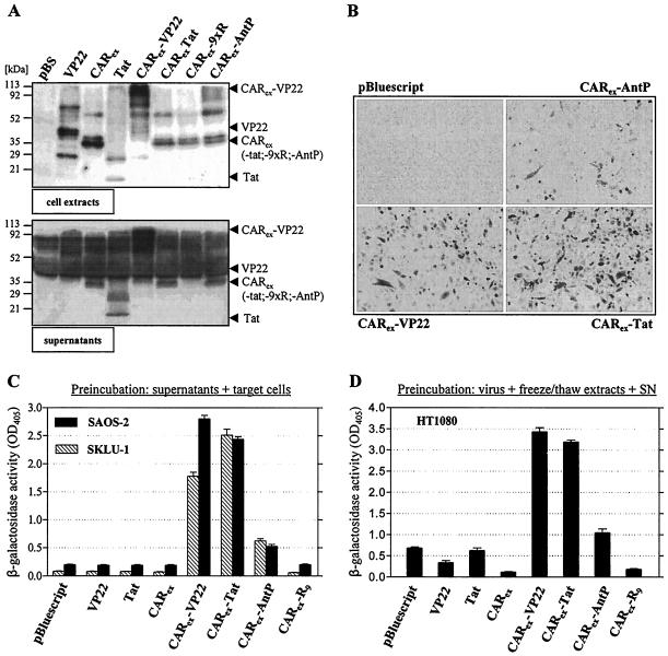
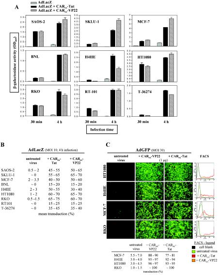
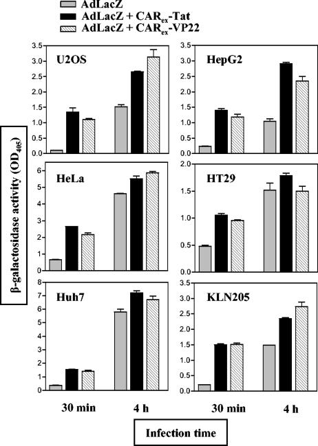
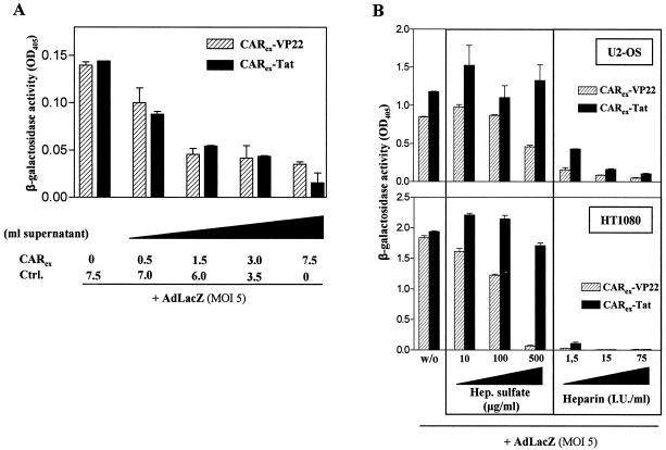
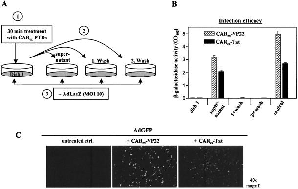
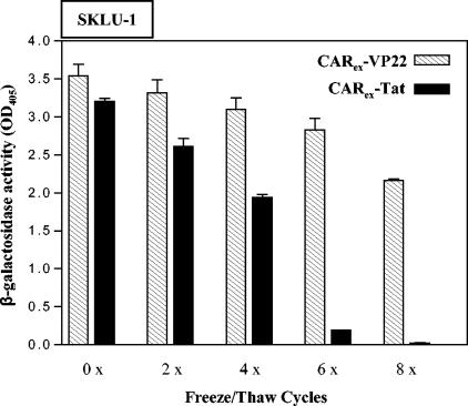
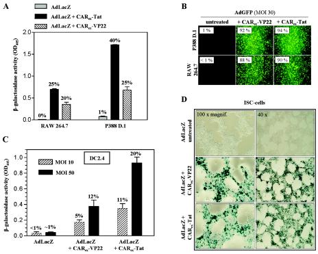
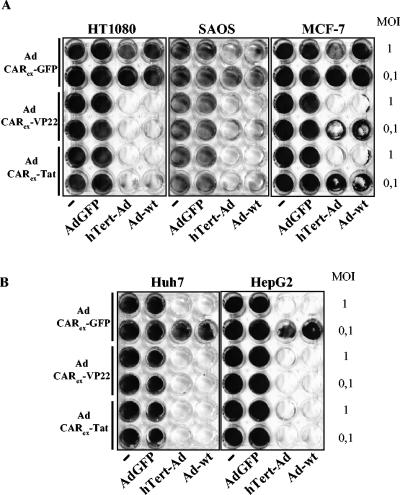
Expression of cellular receptors determines viral tropism and limits gene delivery by viral vectors. Protein transduction domains (PTDs) have been shown to deliver proteins, antisense oligonucleotides, liposomes, or plasmid DNA into cells. In our study, we investigated the role of several PTD motifs in adenoviral infection. When physiologically expressed, a PTD from human immunodeficiency virus transactivator of transcription (Tat) did not improve adenoviral infection. We therefore fused PTDs to the ectodomain of the coxsackievirus-adenovirus receptor (CAR(ex)) to attach PTDs to adenoviral fiber knobs. CAR(ex)-Tat and CAR(ex)-VP22 allowed efficient adenoviral infection in nonpermissive cells and significantly improved viral uptake rates in permissive cells. Dose-dependent competition of CAR(ex)-PTD-mediated infection using CAR(ex) and inhibition experiments with heparin showed that binding of CAR(ex)-PTD to both adenoviral fiber and cellular glycosaminoglycans is essential for the improvement of infection. CAR(ex)-PTD-treated adenoviruses retained their properties after density gradient ultracentrifugation, indicating stable binding of CAR(ex)-PTD to adenoviral particles. Consequently, the mechanism of CAR(ex)-PTD-mediated infection involves coating of the viral fiber knobs by CAR(ex)-PTD, rather than placement of CAR(ex) domains on cell surfaces. Expression of CAR(ex)-PTDs led to enhanced lysis of permissive and nonpermissive tumor cells by replicating adenoviruses, indicating that CAR(ex)-PTDs are valuable tools to improve the efficacy of oncolytic therapy. Together, our study shows that CAR(ex)-PTDs facilitate gene transfer in nonpermissive cells and improve viral uptake at reduced titers and infection times. The data suggest that PTDs fused to virus binding receptors may be a valuable tool to overcome natural tropism of vectors and could be of great interest for gene therapeutic approaches.


Figures

FIG. 1.
FIG. 1.
Plasmid construction overview. Names of the corresponding plasmids are (from top to bottom): pVP22mycHis 2, pCARex-VP22, pCARex-9xArg, pCARex-AntP62-77, pCARex-Tat48-57, pCARex-G/T/S10, and pCAR1-58-Tat48-57.
FIG. 2.
FIG. 2.
Soluble recombinant fusion proteins consisting of the entire ectodomain of hCAR and cell adhesion peptides derived from Tat, VP22, and AntP enable adenoviral infection of SKLU-1, SAOS-2, and HT1080 cells. 293 cells were transfected with expression vectors for CARex-PTDs and control proteins as indicated in the figure. (A) Samples of whole-cell extracts (top) and supernatants (bottom) of producer cells were analyzed for CARex-PTD presence by Western blotting. (B) SKLU-1 cells were treated with equivalent amounts of supernatants from CARex-PTD-transfected 293 cells. AdLacZ was added at an MOI of 10, and the infection was carried out for 4 h. After 48 h, infected SKLU-1 cells were visualized by X-Gal staining. (C) SKLU-1 and SAOS-2 cells were treated as described in the legend to panel B. Infection efficacy was determined by measuring β-galactosidase activity in cell extracts. (D) CARex-PTD-transfected 293 cells were lysed by freeze-thaw cycles. Freeze-thaw lysates were diluted in their respective supernatants, combined with AdLacZ (MOI, 10), incubated for 10 min, and then applied to HT1080 target cells for 4 h. Infection efficacy was determined by a β-galactosidase assay.
FIG. 3.
FIG. 3.
Determination of CARex-VP22- and CARex-Tat-mediated adenoviral infection in nonpermissive or partially permissive tumor cell lines. (A) Different target cell types were overlaid with medium containing purified CARex-VP22 or CARex-Tat at a concentration of 2 nM. AdLacZ (MOI, 10) was then added, and infection was carried out for 30 min or 4 h as indicated. After 48 h of incubation, infection efficacy was determined by measuring β-galactosidase activity in extracts from infected cells. (B) The total degree of infection was determined by X-Gal staining. (C) A total of 1 μg of recombinant protein was dissolved in medium and combined with AdGFP (MOI, 30) and incubated for 10 min prior to a 4-h infection of target cells. After 48 h, GFP expression was monitored by fluorescence microscopy, and infection efficacy was determined by FACS analysis. The results indicate that adenovirus treatment with CARex-VP22 and CARex-Tat facilitates effective infection of nonpermissive cells.
FIG. 4.
FIG. 4.
CARex-VP22 and CARex-Tat improve adenoviral infection of permissive tumor cell lines. Different target cell types were overlaid with medium containing purified CARex-VP22 or CARex-Tat, respectively, at a concentration of 2 nM. AdLacZ (MOI, 10) was then added, and infection was carried out for 30 min or 4 h as indicated. After 48 h of incubation, infection efficacy was determined by β-galactosidase assays of extracts from infected cells.
FIG. 5.
FIG. 5.
CARex-VP22- or CARex-Tat-mediated adenoviral infection can be competed by recombinant CARex lacking a functional PTD and is effectively inhibited by negatively charged heparin and/or heparan sulfate. (A) To block fiber knobs of AdLacZ, the virus was treated for 15 min with various amounts of supernatant from 293 cells expressing CARex. Then, purified CARex-VP22 or CARex-Tat was added at a final concentration of 1 nM, and the mixture was subjected to SKLU-1 cells for an infection period of 30 min. Infected cells were maintained for 48 h and infection efficacy was determined by measuring β-galactosidase activity in cellular extracts. (B) U2-OS (top) and HT1080 (bottom) cells were covered with medium containing 2 nM purified CARex-PTD fusion proteins and increasing amounts of heparan sulfate or heparin, as indicated. AdLacZ (MOI, 5) was added, and infection was carried out for 30 min. Infected cells were maintained for 48 h, and infection efficacy was determined by β-galactosidase assays of cellular extracts.
FIG. 6.
FIG. 6.
CARex-PTD fusion proteins form stable complexes with adenoviral vectors. SKLU-1 cells (dish 1) were treated with CARex fusion proteins (2 nM) and incubated for 30 min. The supernatant and two wash fractions (medium) were then transferred to separate SKLU-1 dishes. For the control sample, the supernatant was left on a dish. Subsequently, all cells were infected with AdLacZ (MOI, 10) for 4 h and incubated for transgene expression. (A) Experimental setup. (B) Results of the corresponding β-galactosidase activity measurements. (C) AdGFP was dialyzed against DMEM and treated with purified recombinant CARex-VP22 or CARex-Tat in a 10-fold molar excess relative to adenoviral fiber knob molecules of AdGFP. As a control, AdGFP received DMEM alone. The virus preparations were then subjected to a CsCl density gradient ultracentrifugation for separation of unbound protein. The virus band was extracted, dialyzed against DMEM, and quantified by determination of optical density at 260 nm. Infection of SKLU-1 cells was carried out for 4 h at an MOI of 50.
FIG. 7.
FIG. 7.
Purified recombinant CARex-VP22 reveals higher stability against thermal influence than CARex-Tat. Purified recombinant CARex-VP22 and CARex-Tat preparations were subjected to freeze-thaw cycles as indicated, mixed with medium, and added to SKLU-1 target cells at a final concentration of 2 nM. Subsequently, cells were infected with AdLacZ (MOI, 10). Infection efficacy was determined by β-galactosidase assays of cellular extracts from infected cells.
FIG. 8.
FIG. 8.
Application of CARex fusion proteins increases adenoviral infection in cell lines derived from immune cells and the nervous system. (A) RAW264.7 macrophages and P388D.1 monocyte/macrophage cells were treated with 2 nM purified recombinant CARex-VP22 or CARex-Tat, respectively, and infected with AdLacZ (MOI of 10; 4 h). Infection efficacy was determined by β-galactosidase assay and X-Gal staining (degree of infection is indicated on top of the bars). (B) AdGFP (MOI, 30) was preincubated with CARex-VP22 or CARex-Tat at a final concentration of 8 nM and then added to RAW264.7 and p388D.1 target cells. Infection efficacy was determined by fluorescence microscopy and FACS. (C) AdLacZ treated with 2 nM CARex-VP22 or CARex-Tat was applied to DC2.4 dendritic cells at MOIs of 10 and 50 for an infection time of 30 min. Infection was quantified by β-galactosidase assay or FACS analysis (AdGFP was applied under the same conditions). (D) Immortalized Schwann (ISC) cells were treated as described in the legend to panel A. The degree of infected ISC cells was visualized by X-Gal staining.
FIG. 9.
FIG. 9.
Receptor-independent infection mediated by CARex-PTD enhances lysis of tumor cell layers by conditionally and nonconditionally replicating Ad vectors. Tumor cell layers were infected with hTERT-Ad, Ad-wt, and Ad-GFP (nonreplicating control) at the MOI indicated. Furthermore, the cells were infected with AdCARex-VP22, AdCARex-Tat, and AdGFP (control) at an MOI of 25 (for low-permissive cell lines SAOS-2, HT1080, and MCF-7) (A) or an MOI of 2 (for permissive cell lines Huh7 and HepG2) (B). After 6 days, replication-associated cell layer destruction was visualized by crystal violet staining.

Similar articles

Cited by

References

    1. Anders, M., R. Hansen, R. X. Ding, K. A. Rauen, M. J. Bissell, and W. M. Korn. 2003. Disruption of 3D tissue integrity facilitates adenovirus infection by deregulating the coxsackievirus and adenovirus receptor. Proc. Natl. Acad. Sci. USA 100:1943-1948. - PMC - PubMed
    1. Bergelson, J. M., J. A. Cunningham, G. Droguett, E. A. Kurt-Jones, A. Krithivas, J. S. Hong, M. S. Horwitz, R. L. Crowell, and R. W. Finberg. 1997. Isolation of a common receptor for coxsackie B viruses and adenoviruses 2 and 5. Science 275:1320-1323. - PubMed
    1. Bergelson, J. M., A. Krithivas, L. Celi, G. Droguett, M. S. Horwitz, T. Wickham, R. L. Crowell, and R. W. Finberg. 1998. The murine CAR homolog is a receptor for coxsackie B viruses and adenoviruses. J. Virol. 72:415-419. - PMC - PubMed
    1. Cohen, C. J., J. T. Shieh, R. J. Pickles, T. Okegawa, J. T. Hsieh, and J. M. Bergelson. 2001. The coxsackievirus and adenovirus receptor is a transmembrane component of the tight junction. Proc. Natl. Acad. Sci. USA 98:15191-15196. - PMC - PubMed
    1. Console, S., C. Marty, C. Garcia-Echeverria, R. Schwendener, and K. Ballmer-Hofer. 2003. Antennapedia and HIV transactivator of transcription (TAT) “protein transduction domains” promote endocytosis of high molecular weight cargo upon binding to cell surface glycosaminoglycans. J. Biol. Chem. 278:35109-35114. - PubMed

Publication types

MeSH terms

LinkOut - more resources