. 2004 Dec;78(24):13793-803.
doi: 10.1128/JVI.78.24.13793-13803.2004.

Replicon system for Lassa virus

Affiliations

Replicon system for Lassa virus

Meike Hass et al. J Virol. 2004 Dec.

Abstract

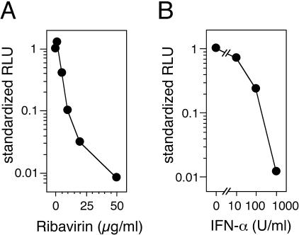
Lassa virus is endemic to West Africa and causes hemorrhagic fever in humans. To facilitate the functional analysis of this virus, a replicon system was developed based on Lassa virus strain AV. Genomic and antigenomic minigenomes (MG) were constructed consisting of the intergenic region of S RNA and a reporter gene (Renilla luciferase) in antisense orientation, flanked by the 5' and 3' untranslated regions of S RNA. MGs were expressed under the control of the T7 promoter. Nucleoprotein (NP), L protein, and Z protein were expressed from plasmids containing the T7 promoter and internal ribosomal entry site. Transfection of cells stably expressing T7 RNA polymerase (BSR T7/5) with MG in the form of DNA or RNA and plasmids for the expression of NP and L protein resulted in high levels of Renilla luciferase expression. The replicon system was optimized with respect to the ratio of the transfected constructs and by modifying the 5' end of the MG. Maximum activity was observed 24 to 36 h after transfection with a signal-to-noise ratio of 2 to 3 log units. Northern blot analysis provided evidence for replication and transcription of the MG. Z protein downregulated replicon activity close to background levels. Treatment with ribavirin and alpha interferon inhibited replicon activity, suggesting that both act on the level of RNA replication, transcription, or ribonucleoprotein assembly. In conclusion, this study describes the first replicon system for a highly pathogenic arenavirus. It is a tool for investigating the mechanisms of replication and transcription of Lassa virus and may facilitate the testing of antivirals outside a biosafety level 4 laboratory.


Figures

FIG. 1.
FIG. 1.
Flowchart of construction of L protein expression plasmid. The L gene was amplified in three fragments by PCR. The primers are indicated above each fragment. Restriction enzyme recognition sites used for cloning are indicated. Sites used for assembly of pUC-L and subcloning of the L gene into pCITE-L are marked by arrows. At the bottom, details of the subcloning of the L gene into the NcoI site of pCITE-L are shown. Note that cloning was possible, although the overhang at the 5′ end of L gene was not fully compatible with an NcoI overhang.
FIG. 2.
FIG. 2.
Constructs used for establishment of Lassa virus minireplicon. (A) Plasmids for expression of Lassa virus strain AV proteins. (B) Types of MGs based on Lassa virus S RNA. DNA is indicated by a straight line, and RNA is indicated by a wavy line. Functional elements are abbreviated as follows: T7p, T7 promoter; IRES, internal ribosomal entry site; UTR, untranslated region with conserved termini; IGR, intergenic region; HDR, hepatitis delta ribozyme; T7t, T7 transcriptional terminator; luc, luciferase.
FIG. 3.
FIG. 3.
Immunoblot analysis of plasmid-expressed Lassa virus proteins. MVA-T7-infected BHK-21 cells were transfected with the constructs as indicated above the blots. Alternatively, Vero cells were infected with Lassa virus (only panel B). Negative control cells (NC) were infected with MVA-T7 and mock transfected. The background bands in the NC lane of panel C result from an overloading of the gel. The blots were incubated with the following antibodies: rabbit anti-Lassa Z protein (A), rabbit anti-Lassa NP (B), and anti-FLAG M2 monoclonal antibody (C and D).
FIG. 4.
FIG. 4.
Influence of nontemplated nucleotides at the 5′ end of Lassa virus MGs on replicon activity. Experiments were performed with runoff MGs based on genomic (A) and antigenomic (B) Lassa virus S RNA. The upper panels show schematic drawings of the MG variants tested. The variants were generated by mutagenic PCR and named according to the number of nontemplated G residues at the 5′ end. Functional elements are abbreviated as follows: UTR, untranslated region with conserved termini; IGR, intergenic region; Ren, Renilla luciferase. The lower panels show the replicon activity depending on the presence (+) or absence (−) of NP or L protein and the type of MG. BSR T7/5 cells (5 × 10⁵ cells per well of a six-well plate) were transfected with 2 μg of MG, 1.5 μg of pCITE-NP, and/or 0.5 μg of pCITE-L. Empty pCITE-2 was used to keep the overall DNA amount constant if pCITE-NP or pCITE-L was not included in the transfection mixture. MG variants were transfected as either DNA (PCR fragment) or RNA (in vitro transcript). Ren-Luc activity was measured in RLU 24 h posttransfection. Note the logarithmic scale of the diagrams.
FIG. 5.
FIG. 5.
Determination of the optimal ratios between MG, pCITE-NP, and pCITE-L. (A) Titration of ratio between pCITE-NP and pCITE-L. BSR T7/5 cells (5 × 10⁵ cells per well of a six-well plate) were transfected with 2 μg of genomic MG DNA (PCR fragment of construct 1G in Fig. 4A) and a total amount of 2 μg of pCITE-L and pCITE-NP in different ratios (filled squares). In negative-control experiments, pCITE-L was replaced by empty pCITE-2 (open circles). Ren-Luc activity was measured in RLU 24 h posttransfection. (B) Titration of ratio between MG and protein expression plasmids. BSR T7/5 cells (105 cells per well of a 24-well plate) were transfected with MG and a 1:1 mixture of pCITE-NP and pCITE-L (filled squares). The ratio of MG to pCITE-NP (identical to the ratio of MG to pCITE-L) differed in each experiment, but the total amount of transfected DNA was kept constant at 800 ng. In negative-control experiments, MG DNA was replaced by empty pCITE-2 (open circles).
FIG. 6.
FIG. 6.
Time kinetics of replicon activity. (A) Time kinetics with MG DNA. BSR T7/5 cells (105 cells per well of a 24-well plate) were transfected with 270 ng of genomic MG DNA (PCR fragment of construct 1G in Fig. 4A), 270 ng of pCITE-NP, 270 ng of pCITE-L, and 10 ng of pCITE-FF-luc. Cells were lysed at different times posttransfection, and Renilla (filled squares) and firefly luciferase activities (open circles) were measured in RLU. In negative-control experiments, pCITE-L was replaced by empty pCITE-2 (filled triangles). (B) Time kinetics with MG RNA. Experiments were done as described above except that 270 ng of genomic MG RNA (in vitro transcript of construct 1G in Fig. 4A) was used.
FIG. 7.
FIG. 7.
Northern blot analysis of Lassa virus replicon. (A) Detection of antigenomic RNA/Ren-Luc mRNA. BSR T7/5 cells (1.2 × 10⁶ cells per 50-mm-diameter dish) were transfected with 2.7 μg of genomic MG DNA (PCR fragment of construct 1G in Fig. 4A), 2.7 μg of pCITE-NP, 2.7 μg of pCITE-L, and 100 ng of pCITE-FF-luc (lanes +). In control experiments, pCITE-L was replaced by empty pCITE-2 (lanes −). Total RNA was isolated at different time posttransfection, and Northern blot hybridization was performed with a 32P-labeled antisense probe of the Ren-Luc gene. The insert in the lower left corner shows the signal area of lanes 0 h and 6 h with enhanced contrast. The methylene blue-stained 28S rRNA is shown below the blot as a semiquantitative marker for gel loading and RNA transfer. (B) Detection of genomic RNA. Experiments were done as described above except that a 32P-labeled sense probe of the Ren-Luc gene was used for Northern blot hybridization. (C) Quantification of the RNA signals in panels A (lanes +, filled squares) and B (lanes +, open squares; lanes −, open circles). Arrows indicate the fraction of genomic RNA resulting from MG replication by L protein.
FIG. 8.
FIG. 8.
Inhibitory effect of Lassa virus Z protein on minireplicon activity. BSR T7/5 cells (105 cells per well of a 24-well plate) were transfected with 200 ng of genomic MG DNA (PCR fragment of construct 1G in Fig. 4A), 200 ng of pCITE-NP, 200 ng of pCITE-L, and 10 ng of pCITE-FF-luc. Various quantities of pCITE-Z were cotransfected. The total amount of transfected DNA was kept constant by the addition of pCITE-2. Cells were lysed 24 h posttransfection, and Renilla (filled squares) and firefly luciferase activities (open circles) were measured in RLU. Note the logarithmic scale of the diagram.
FIG. 9.
FIG. 9.
Inhibition of replicon activity by ribavirin and IFN-α. (A) Effect of ribavirin. BSR T7/5 cells (105 cells per well of a 24-well plate) were transfected with 270 ng of genomic MG DNA (PCR fragment of construct 1G in Fig. 4A), 270 ng of pCITE-NP, 270 ng of pCITE-L, and 10 ng of pCITE-FF-luc. Different concentrations of ribavirin were added 4 h after transfection. Cells were lysed 24 h posttransfection, and Renilla and firefly luciferase activities were measured. Ren-Luc levels were corrected with the firefly luciferase levels (standardized RLU). The number of RLU in untreated cells was defined as 1. (B) Effect of IFN-α. Experiments were done as described above except that cells were treated with human IFN-α for 24 h before transfection. The same concentrations were added again 4 h after transfection. Note the logarithmic scale of the diagrams.

Similar articles

Cited by

References

    1. Andrei, G., and E. De Clercq. 1990. Inhibitory effect of selected antiviral compounds on arenavirus replication in vitro. Antivir. Res. 14:287-299. - PubMed
    1. Asper, M., T. Sternsdorf, M. Hass, C. Drosten, A. Rhode, H. Schmitz, and S. Günther. 2004. Inhibition of different Lassa virus strains by alpha and gamma interferons and comparison with a less pathogenic arenavirus. J. Virol. 78:3162-3169. - PMC - PubMed
    1. Auperin, D. D., D. R. Sasso, and J. B. McCormick. 1986. Nucleotide sequence of the glycoprotein gene and intergenic region of the Lassa virus S genome RNA. Virology 154:155-167. - PubMed
    1. Auperin, D. D., V. Romanowski, M. Galinski, and D. H. Bishop. 1984. Sequencing studies of pichinde arenavirus S RNA indicate a novel coding strategy, an ambisense viral S RNA. J. Virol. 52:897-904. - PMC - PubMed
    1. Bausch, D. G., A. H. Demby, M. Coulibaly, J. Kanu, A. Goba, A. Bah, N. Conde, H. L. Wurtzel, K. F. Cavallaro, E. Lloyd, F. B. Baldet, S. D. Cisse, D. Fofona, I. K. Savane, R. T. Tolno, B. Mahy, K. D. Wagoner, T. G. Ksiazek, C. J. Peters, and P. E. Rollin. 2001. Lassa fever in Guinea. I. Epidemiology of human disease and clinical observations. Vector Borne Zoonotic Dis. 1:269-281. - PubMed

Publication types

Associated data

LinkOut - more resources