Recombinant infectious bronchitis coronavirus Beaudette with the spike protein gene of the pathogenic M41 strain remains attenuated but induces protective immunity
- PMID: 15564488
- PMCID: PMC533908
- DOI: 10.1128/JVI.78.24.13804-13811.2004
Recombinant infectious bronchitis coronavirus Beaudette with the spike protein gene of the pathogenic M41 strain remains attenuated but induces protective immunity
Abstract
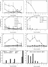
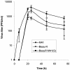
We have replaced the ectodomain of the spike (S) protein of the Beaudette strain (Beau-R; apathogenic for Gallus domesticus chickens) of avian infectious bronchitis coronavirus (IBV) with that from the pathogenic M41 strain to produce recombinant IBV BeauR-M41(S). We have previously shown that this changed the tropism of the virus in vitro (R. Casais, B. Dove, D. Cavanagh, and P. Britton, J. Virol. 77:9084-9089, 2003). Herein we have assessed the pathogenicity and immunogenicity of BeauR-M41(S). There were no consistent differences in pathogenicity between the recombinant BeauR-M41(S) and its apathogenic parent Beau-R (based on snicking, nasal discharge, wheezing, watery eyes, rales, and ciliostasis in trachea), and both replicated poorly in trachea and nose compared to M41; the S protein from the pathogenic M41 had not altered the apathogenic nature of Beau-R. Both Beau-R and BeauR-M41(S) induced protection against challenge with M41 as assessed by absence of recovery of challenge virus and nasal exudate. With regard to snicking and ciliostasis, BeauR-M41(S) induced greater protection (seven out of nine chicks [77%]; assessed by ciliostasis) than Beau-R (one out of nine; 11%) but less than M41 (100%). The greater protection induced by BeauR-M41(S) against M41 may be related to the ectodomain of the spike protein of Beau-R differing from that of M41 by 4.1%; a small number of epitopes on the S protein may play a disproportionate role in the induction of immunity. The results are promising for the prospects of S-gene exchange for IBV vaccine development.
Figures


Similar articles
-
Recombinant Infectious Bronchitis Viruses Expressing Chimeric Spike Glycoproteins Induce Partial Protective Immunity against Homologous Challenge despite Limited Replication In Vivo.J Virol. 2018 Nov 12;92(23):e01473-18. doi: 10.1128/JVI.01473-18. Print 2018 Dec 1. J Virol. 2018. PMID: 30209177 Free PMC article.
-
Safety and efficacy of an infectious bronchitis virus used for chicken embryo vaccination.Vaccine. 2006 Nov 17;24(47-48):6830-8. doi: 10.1016/j.vaccine.2006.06.040. Epub 2006 Jul 5. Vaccine. 2006. PMID: 16860445 Free PMC article.
-
A recombinant avian infectious bronchitis virus expressing a heterologous spike gene belonging to the 4/91 serotype.PLoS One. 2011;6(8):e24352. doi: 10.1371/journal.pone.0024352. Epub 2011 Aug 30. PLoS One. 2011. PMID: 21912629 Free PMC article.
-
Recombinant avian infectious bronchitis virus expressing a heterologous spike gene demonstrates that the spike protein is a determinant of cell tropism.J Virol. 2003 Aug;77(16):9084-9. doi: 10.1128/jvi.77.16.9084-9089.2003. J Virol. 2003. PMID: 12885925 Free PMC article.
-
Anatomical Poems about the Breast ("Le Beau Tetin") and Anatomical Proportions.Plast Reconstr Surg Glob Open. 2021 Jun 24;9(6):e3652. doi: 10.1097/GOX.0000000000003652. eCollection 2021 Jun. Plast Reconstr Surg Glob Open. 2021. PMID: 34235037 Free PMC article. Review. No abstract available.
Cited by
-
First evidence of the emergence of novel putative infectious bronchitis virus genotypes in Cuba.Res Vet Sci. 2012 Oct;93(2):1046-9. doi: 10.1016/j.rvsc.2012.01.012. Epub 2012 Feb 21. Res Vet Sci. 2012. PMID: 22357366 Free PMC article.
-
The spike protein of the apathogenic Beaudette strain of avian coronavirus can elicit a protective immune response against a virulent M41 challenge.PLoS One. 2024 Jan 24;19(1):e0297516. doi: 10.1371/journal.pone.0297516. eCollection 2024. PLoS One. 2024. PMID: 38265985 Free PMC article.
-
Molecular Characterization of Infectious Bronchitis Virus Strain HH06 Isolated in a Poultry Farm in Northeastern China.Front Vet Sci. 2021 Dec 16;8:794228. doi: 10.3389/fvets.2021.794228. eCollection 2021. Front Vet Sci. 2021. PMID: 34977225 Free PMC article.
-
Binding of avian coronavirus spike proteins to host factors reflects virus tropism and pathogenicity.J Virol. 2011 Sep;85(17):8903-12. doi: 10.1128/JVI.05112-11. Epub 2011 Jun 22. J Virol. 2011. PMID: 21697468 Free PMC article.
-
Activation of the chicken type I interferon response by infectious bronchitis coronavirus.J Virol. 2015 Jan 15;89(2):1156-67. doi: 10.1128/JVI.02671-14. Epub 2014 Nov 5. J Virol. 2015. PMID: 25378498 Free PMC article.
References
-
- Adzhar, A., R. E. Gough, D. Haydon, K. Shaw, P. Britton, and D. Cavanagh. 1997. Molecular analysis of the 793/B serotype of infectious bronchitis virus in Great Britain. Avian Pathol. 26:625-640. - PubMed
-
- Binns, M. M., M. E. G. Boursnell, F. M. Tomley, and T. D. K. Brown. 1986. Comparison of the spike precursor sequences of coronavirus IBV strains M41 and 6/82 with that of IBV Beaudette. J. Gen. Virol. 67:2825-2831. - PubMed
-
- Butler, M., and W. J. Ellaway. 1972. Comparative studies on the infectivity of avian respiratory viruses for eggs, cell cultures and tracheal explants. J. Comp. Pathol. 82:327-332. - PubMed
Publication types
MeSH terms
Substances
LinkOut - more resources
Full Text Sources
Other Literature Sources

