. 2004 Dec;78(24):13865-70.
doi: 10.1128/JVI.78.24.13865-13870.2004.

Prototype foamy virus envelope glycoprotein leader peptide processing is mediated by a furin-like cellular protease, but cleavage is not essential for viral infectivity

Affiliations

Prototype foamy virus envelope glycoprotein leader peptide processing is mediated by a furin-like cellular protease, but cleavage is not essential for viral infectivity

Anja Duda et al. J Virol. 2004 Dec.

Abstract

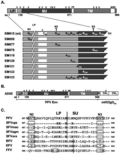
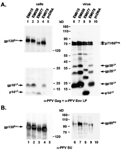
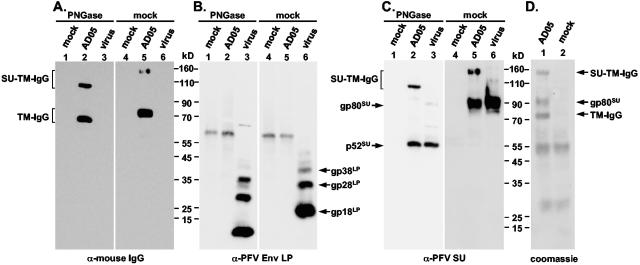
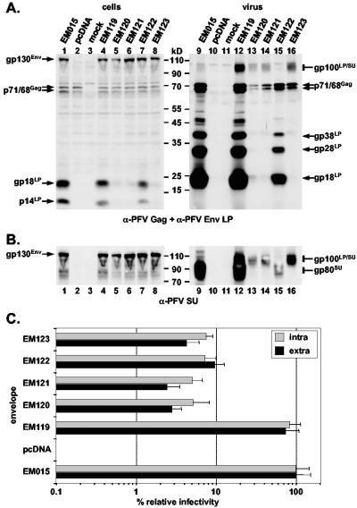
Analogous to cellular glycoproteins, viral envelope proteins contain N-terminal signal sequences responsible for targeting them to the secretory pathway. The prototype foamy virus (PFV) envelope (Env) shows a highly unusual biosynthesis. Its precursor protein has a type III membrane topology with both the N and C terminus located in the cytoplasm. Coexpression of FV glycoprotein and interaction of its leader peptide (LP) with the viral capsid is essential for viral particle budding and egress. Processing of PFV Env into the particle-associated LP, surface (SU), and transmembrane (TM) subunits occur posttranslationally during transport to the cell surface by yet-unidentified cellular proteases. Here we provide strong evidence that furin itself or a furin-like protease and not the signal peptidase complex is responsible for both processing events. N-terminal protein sequencing of the SU and TM subunits of purified PFV Env-immunoglobulin G immunoadhesin identified furin consensus sequences upstream of both cleavage sites. Mutagenesis analysis of two overlapping furin consensus sequences at the PFV LP/SU cleavage site in the wild-type protein confirmed the sequencing data and demonstrated utilization of only the first site. Fully processed SU was almost completely absent in viral particles of mutants having conserved arginine residues replaced by alanines in the first furin consensus sequence, but normal processing was observed upon mutation of the second motif. Although these mutants displayed a significant loss in infectivity as a result of reduced particle release, no correlation to processing inhibition was observed, since another mutant having normal LP/SU processing had a similar defect.


Figures

FIG. 1.
FIG. 1.
Schematic illustration of the PFV Env proteins. (A) Schematic outline of the PFV Env domain organization. The LP-SU region is enlarged below the full-length precursor protein and altered residues of the individual mutants are indicated in comparison to the wild-type sequence. (B) Schematic outline of the PFV Env immunoadhesin domain organization. (C) Sequence alignment of the LP/SU cleavage site and adjacent regions. The arginine residues of the minimal furin cleavage site consensus sequences are in bold letters. A solid black line indicates the boundaries of the PFV LP and SU subunits experimentally determined in this study. A dotted black line indicates the potential boundaries of the glycoproteins of other FV species. N glycosylation sites are boxed. Identical amino acids, except arginine residues, are indicated by hyphens. nsrsid9847724\delrsid9847724 FP, fusion peptide; MSD, membrane-spanning domain; h, hydrophobic domain of the leader peptide; mHCIgG2a, mouse IgG2a heavy-chain constant region; H, hinge region; CH2 or CH3, constant Ig heavy-chain domain 2 or 3; N1 to N3, potential N glycosylation site 1 to 3; N-linked carbohydrate chains are indicated as Y. GenBank accession numbers for the cited viral genomes are U21247 for PFV, U04327 for simian FV-chimpanzee (SFVcpz), X54482 for simian FV-macaque (SFVmac), M74895 for simian FV-African green monkey (SFVagm), AJ544579 for simian FV-orangutan (SFVora), U94514 for bovine FV (BFV), AF201902 for equine FV (EFV), and U85043 for FFV.
FIG. 2.
FIG. 2.
Analysis of PFV Env N glycosylation mutants. Western blot analysis of 293T cell (cells) and purified PFV particle (virus) lysates using (A) anti-PFV Gag- and anti-PFV Env LP (aa 1 to 86)-specific polyclonal rabbit antisera or (B) anti-PFV-SU (P3E10) monoclonal hybridoma supernatant. The individual PFV proteins are indicated. 293T cells were cotransfected with the PFV Gag/Pol-expressing, replication-defective retroviral vector pczDWP001 and the respective PFV Env expression construct as indicated. Lanes 1 to 5, cell lysates; lanes 6 to 10, the corresponding viral particle lysates.
FIG. 3.
FIG. 3.
Characterization of a PFV Env-mIgG2a immunoadhesin. The AD05 PFV Env-IgG (AD05) immunoadhesin was purified in small (A-C) or medium scale (D) by protein A precipitation from transfected 293T cell supernatant. As control, protein A precipitate from equal amounts of supernatant from 239T transfected with the empty expression vector pczCFG5IEGZ (mock) was used. Purified PFV particles (virus) were harvested by ultracentrifugation through 20% sucrose from the supernatant of 293T cells cotransfected with pczDWP001 and pczHFVenvEM015 expression constructs. Prior to separation by sodium dodecyl sulfate-polyacrylamide gel electrophoresis, samples were deglycosylated using PNGase F (PNGase) or mock (mock) incubated. After transfer of the proteins to polyvinylidene difluoride membranes, they were analyzed by Western blot using (A) anti-mouse IgG-, (B) anti-PFV Env LP-, and (C) anti-PFV Env SU-specific antibodies or directly Coomassie stained (D). The individual PFV proteins are indicated on the sides of the autoradiograms.
FIG. 4.
FIG. 4.
Characterization of potential PFV Env furin cleavage site mutants. Western blot analysis of 293T cell (cells) and purified PFV particle (virus) lysates using (A) anti-PFV Gag- and anti-PFV Env LP (aa1 to 86)-specific polyclonal rabbit antisera or (B) anti-PFV-SU (P3E10) monoclonal hybridoma supernatant. The individual PFV proteins are indicated. 293T cells were cotransfected with the PFV Gag/Pol-expressing, replication-defective retroviral vector pczDWP001 and the respective PFV Env expression construct as indicated. Lanes 1 to 8, cell lysates; lanes 9 to 16, the corresponding viral particle lysates; mock, lysate from cells transfected only with pcDNA vector. (C) Relative infectivity of extracellular (black bars) and intracellular (grey bars) PFV particles pseudotyped with the individual PFV Env proteins as indicated on the left in comparison to the wild type. Mean values and standard deviations of two independent experiments with a total of 12 values for each individual Env protein are shown.

References

    1. Bansal, A., K. L. Shaw, B. H. Edwards, P. A. Goepfert, and M. J. Mulligan. 2000. Characterization of the R572T point mutant of a putative cleavage site in human foamy virus Env. J. Virol. 74:2949-2954. - PMC - PubMed
    1. Baunach, G., B. Maurer, H. Hahn, M. Kranz, and A. Rethwilm. 1993. Functional analysis of human foamy virus accessory reading frames. J. Virol. 67:5411-5418. - PMC - PubMed
    1. Dinev, D., B. W. Jordan, B. Neufeld, J. D. Lee, D. Lindemann, U. R. Rapp, and S. Ludwig. 2001. Extracellular signal regulated kinase 5 (ERK5) is required for the differentiation of muscle cells. EMBO Rep. 2:829-834. - PMC - PubMed
    1. Du Bridge, R. B., P. Tang, H. C. Hsia, P. M. Leong, J. H. Miller, and M. P. Calos. 1987. Analysis of mutation in human cells by using an Epstein-Barr virus shuttle system. Mol. Cell. Biol. 7:379-387. - PMC - PubMed
    1. Feldmann, H., V. E. Volchkov, V. A. Volchkova, U. Stroher, and H. D. Klenk. 2001. Biosynthesis and role of filoviral glycoproteins. J. Gen. Virol. 82:2839-2848. - PubMed

Publication types

LinkOut - more resources