. 2004 Dec;78(24):13891-900.
doi: 10.1128/JVI.78.24.13891-13900.2004.

Experimental infection of NOD/SCID mice reconstituted with human CD34+ cells with Epstein-Barr virus

Affiliations

Experimental infection of NOD/SCID mice reconstituted with human CD34+ cells with Epstein-Barr virus

Miguel Islas-Ohlmayer et al. J Virol. 2004 Dec.

Abstract

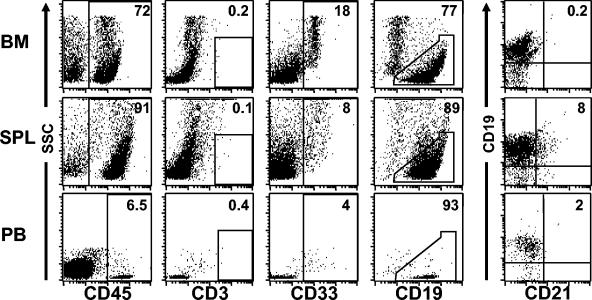
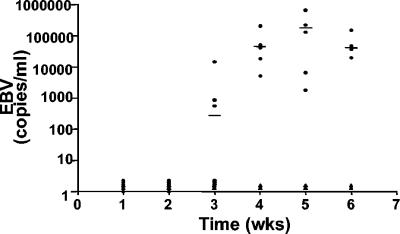
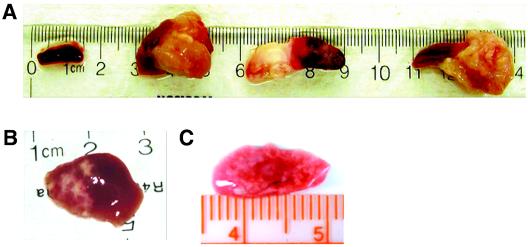
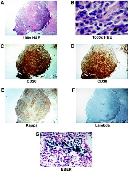
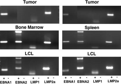
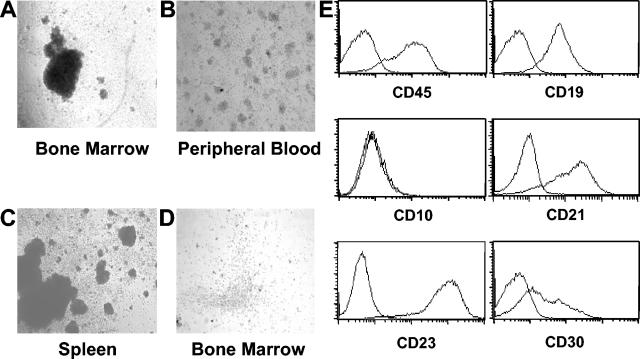
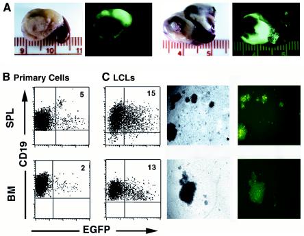
Epstein-Barr virus (EBV)-induced lymphoproliferative disease is an important complication in the context of immune deficiency. Impaired T-cell immunity allows the outgrowth of transformed cells with the subsequent production of predominantly B-cell lymphomas. Currently there is no in vivo model that can adequately recapitulate EBV infection and its association with B-cell lymphomas. NOD/SCID mice engrafted with human CD34(+) cells and reconstituted mainly with human B lymphocytes may serve as a useful xenograft model to study EBV infection and pathogenesis. We therefore infected reconstituted mice with EBV. High levels of viral DNA were detected in the peripheral blood of all infected mice. All infected mice lost weight and showed decreased activity levels. Infected mice presented large visible tumors in multiple organs, most prominently in the spleen. These tumors stained positive for human CD79a, CD20, CD30, and EBV-encoded RNAs and were light chain restricted. Their characterization is consistent with that of large cell immunoblastic lymphoma. In addition, tumor cells expressed EBNA1, LMP1, and LMP2a mRNAs, which is consistent with a type II latency program. EBV(+) lymphoblastoid cell lines expressing human CD45, CD19, CD21, CD23, CD5, and CD30 were readily established from the bone marrow and spleens of infected animals. Finally, we also demonstrate that infection with an enhanced green fluorescent protein (EGFP)-tagged virus can be monitored by the detection of infected EGFP(+) cells and EGFP(+) tumors. These data demonstrate that NOD/SCID mice that are reconstituted with human CD34(+) cells are susceptible to infection by EBV and accurately recapitulate important aspects of EBV pathogenesis.


Figures

FIG. 1.
FIG. 1.
Engraftment of preconditioned NOD/SCID mice with human CD34+ cells results predominantly in the production of human B cells. A flow cytometry analysis of human cells (CD45+) that were present in the bone marrow, spleens, and peripheral blood of reconstituted mice showed the presence of abundant CD19+ human B cells in all hematopoietic tissues. Consistent with previous reports, human CD33+ myeloid cells, but no human T cells (CD3+), were detected in these mice in addition to B cells. CD21+ B cells were only present in the spleen. Note that the analysis of CD19, CD33, CD3, and CD21 expression was performed on cells gated through hCD45. Data are from one representative mouse of a group of eight mice.
FIG. 2.
FIG. 2.
Analysis of EBV loads in peripheral blood after infection of NOD/SCID mice that were reconstituted with human CD34+ cells. DNAs were extracted from blood samples collected at weekly intervals from infected and control (transplant recipients that were not infected) mice. Samples were analyzed for the presence of EBV DNA by real-time PCR. Note that no virus was detected in any of the mice during the first 2 weeks postinfection and that by 4 weeks postinfection all animals had high levels of viral DNA in their blood. •, infected mice; ▴, control uninfected mice.
FIG. 3.
FIG. 3.
Development of tumors after infection with EBV in NOD/SCID mice that were reconstituted with human CD34+ cells. At 5 to 6 weeks postinfection, all infected animals had developed tumors in their spleens. (A) Spleen from a control (not infected) reconstituted mouse and spleens from three different EBV-infected mice (n = 7). Disseminated disease was also seen in other organs, including livers (B) and lungs (C).
FIG. 4.
FIG. 4.
Histological characterization of EBV-induced tumors in infected NOD/SCID mice. (A and B) Hematoxylin and eosin (H&E) staining of a representative tumor originating from the spleen of an infected mouse (magnification, ×100 and ×1,000, respectively). (C to F) Immunohistochemical staining with antibodies to human CD20 and CD30 and with antibodies to human immunoglobulin kappa or lambda chains. (G) In situ staining for EBERs in a section from the same tumor.
FIG. 5.
FIG. 5.
Latency analysis of EBV in tumor, bone marrow, and spleen cells and in vitro-expanded LCLs. RT-PCR analyses of EBV gene expression were performed with RNAs obtained from tumor cells isolated from two different infected mice (top), from spleen and bone marrow cells (middle), and from LCLs expanded in vitro from bone marrow and spleen cultures (bottom). The PCR products shown correspond to EBNA1, EBNA2, LMP1, and LMP2a. The plus and minus signs indicate whether or not the RNA samples were reverse transcribed prior to PCR amplification. Note the absence of the PCR amplification product for EBNA2 in the two tumor samples and its presence in the bone marrow, spleen, and LCL samples (all were amplified with the same sets of primers and under identical conditions).
FIG. 6.
FIG. 6.
Flow cytometric analysis of LCLs derived from EBV-infected mice. (A to C) In vitro-cultured cells obtained from the bone marrow, peripheral blood, and spleens of infected mice resulted in the outgrowth of EBV+ LCLs. (D) In contrast, note the absence of cell clumps from a bone marrow sample obtained from a mouse that was a transplant recipient of human CD34+ cells but was not infected with EBV. (E) Flow cytometric analysis of in vitro-expanded LCLs for cell surface expression of human CD45, CD19, CD10, CD21, CD23, and CD30.
FIG. 7.
FIG. 7.
In situ analysis of EGFP expression in tumors resulting from infection with EBV tagged with EGFP. (A) Infected mice were sacrificed at 5 to 7 weeks postinfection, and internal organs from two different mice with visible tumors were removed for analysis under UV light with a dissecting microscope. (B) EGFP-expressing human B cells were also detected in the spleens (SPL) (not tumor tissue) and bone marrow (BM) of these mice (left dot plots and pictures). (C) LCLs obtained after in vitro culturing of cells from spleens or peripheral blood also expressed EGFP (right dot plots and images).

References

    1. Aljurf, M. D., T. W. Owaidah, A. Ezzat, E. Ibrahim, and A. Tbakhi. 2003. Antigen- and/or immune-driven lymphoproliferative disorders. Ann. Oncol. 14:1595-1606. - PubMed
    1. Babcock, G. J., and D. A. Thorley-Lawson. 2000. Tonsillar memory B cells, latently infected with Epstein-Barr virus, express the restricted pattern of latent genes previously found only in Epstein-Barr virus-associated tumors. Proc. Natl. Acad. Sci. USA 97:12250-12255. - PMC - PubMed
    1. Baumforth, K. R., L. S. Young, K. J. Flavell, C. Constandinou, and P. G. Murray. 1999. The Epstein-Barr virus and its association with human cancers. Mol. Pathol. 52:307-322. - PMC - PubMed
    1. Bollard, C. M., B. Savoldo, C. M. Rooney, and H. E. Heslop. 2003. Adoptive T-cell therapy for EBV-associated post-transplant lymphoproliferative disease. Acta Haematologica 110:139-148. - PubMed
    1. Chang, Y., G. C. Bosma, and M. J. Bosma. 1995. Development of B cells in scid mice with immunoglobulin transgenes: implications for the control of V(D)J recombination. Immunity 2:607-616. - PubMed

Publication types

MeSH terms